US8993245B2 - Biosensor for detecting multiple epitopes on a target - Google Patents
Biosensor for detecting multiple epitopes on a target Download PDFInfo
- Publication number
- US8993245B2 US8993245B2 US13/133,198 US200913133198A US8993245B2 US 8993245 B2 US8993245 B2 US 8993245B2 US 200913133198 A US200913133198 A US 200913133198A US 8993245 B2 US8993245 B2 US 8993245B2
- Authority
- US
- United States
- Prior art keywords
- target
- complementary
- epitope
- length
- binding agent
- Prior art date
- Legal status (The legal status is an assumption and is not a legal conclusion. Google has not performed a legal analysis and makes no representation as to the accuracy of the status listed.)
- Expired - Fee Related
Links
Images


Classifications
-
- G—PHYSICS
- G01—MEASURING; TESTING
- G01N—INVESTIGATING OR ANALYSING MATERIALS BY DETERMINING THEIR CHEMICAL OR PHYSICAL PROPERTIES
- G01N33/00—Investigating or analysing materials by specific methods not covered by groups G01N1/00 - G01N31/00
- G01N33/48—Biological material, e.g. blood, urine; Haemocytometers
- G01N33/50—Chemical analysis of biological material, e.g. blood, urine; Testing involving biospecific ligand binding methods; Immunological testing
- G01N33/53—Immunoassay; Biospecific binding assay; Materials therefor
- G01N33/569—Immunoassay; Biospecific binding assay; Materials therefor for microorganisms, e.g. protozoa, bacteria, viruses
- G01N33/56911—Bacteria
- G01N33/56916—Enterobacteria, e.g. shigella, salmonella, klebsiella, serratia
-
- G—PHYSICS
- G01—MEASURING; TESTING
- G01N—INVESTIGATING OR ANALYSING MATERIALS BY DETERMINING THEIR CHEMICAL OR PHYSICAL PROPERTIES
- G01N33/00—Investigating or analysing materials by specific methods not covered by groups G01N1/00 - G01N31/00
- G01N33/48—Biological material, e.g. blood, urine; Haemocytometers
- G01N33/50—Chemical analysis of biological material, e.g. blood, urine; Testing involving biospecific ligand binding methods; Immunological testing
- G01N33/53—Immunoassay; Biospecific binding assay; Materials therefor
- G01N33/536—Immunoassay; Biospecific binding assay; Materials therefor with immune complex formed in liquid phase
- G01N33/542—Immunoassay; Biospecific binding assay; Materials therefor with immune complex formed in liquid phase with steric inhibition or signal modification, e.g. fluorescent quenching
-
- G—PHYSICS
- G01—MEASURING; TESTING
- G01N—INVESTIGATING OR ANALYSING MATERIALS BY DETERMINING THEIR CHEMICAL OR PHYSICAL PROPERTIES
- G01N2333/00—Assays involving biological materials from specific organisms or of a specific nature
- G01N2333/195—Assays involving biological materials from specific organisms or of a specific nature from bacteria
- G01N2333/24—Assays involving biological materials from specific organisms or of a specific nature from bacteria from Enterobacteriaceae (F), e.g. Citrobacter, Serratia, Proteus, Providencia, Morganella, Yersinia
- G01N2333/245—Escherichia (G)
-
- G—PHYSICS
- G01—MEASURING; TESTING
- G01N—INVESTIGATING OR ANALYSING MATERIALS BY DETERMINING THEIR CHEMICAL OR PHYSICAL PROPERTIES
- G01N2333/00—Assays involving biological materials from specific organisms or of a specific nature
- G01N2333/195—Assays involving biological materials from specific organisms or of a specific nature from bacteria
- G01N2333/24—Assays involving biological materials from specific organisms or of a specific nature from bacteria from Enterobacteriaceae (F), e.g. Citrobacter, Serratia, Proteus, Providencia, Morganella, Yersinia
- G01N2333/255—Salmonella (G)
Definitions
- Pathogenic bacteria are responsible for over fifty infectious diseases. Methodologies allowing detection of the pathogens in clinical, food, water and environmental samples are an important component of infectious disease diagnosis, treatment and prevention.
- pathogen detection involves traditional methods based on cell culture and colony counting, antigen detection methods, PCR-based methods, and various biosensors. Each of these methods has its own strengths and weaknesses. Traditional methods are robust and sensitive, but very slow. Antigen and PCR-based methods are much faster but are technically demanding and in the case of PCR-based methods, prone to false-positives. Biosensors offer the promise of much shorter detection times but require more development before they become a real alternative. There is clearly a need for new detection methodologies that would overcome the limitations of currently existing technologies.
- One aspect of the present invention encompasses a method for detecting a target comprising at least one repeating epitope.
- the method comprises contacting a sample comprising the target with a molecular biosensor.
- the biosensor comprises: R1-R2-R3-R4; and R5-R6-R7-R8;
- FIG. 1 depicts (A) the design of a molecular biosensor.
- ANTI 1 and ANTI 2 depict antibodies labeled with signaling oligonucleotides. T corresponds to the target protein.
- B Proof-of-principle for molecular biosensors.
- Black line emission spectrum of 20 nM ANTI 1 labeled with fluorescein.
- Red line Emission spectrum of a mixture of 20 nM ANTI 1 and 25 nM ANTI 2 labeled with Cy5.
- Blue line 20 nM ANTI 1 and 25 nM ANTI 2 in the presence of 20 nM human cardiac troponin I. Excitation was at 490 nm.
- Inset FRET signals for each sample (emission at 670 nm with the excitation at 490 nm).
- C Cardiac troponin sensor response at low concentrations of the protein.
- FIG. 2 depicts the design of antibody-based sensor for detecting pathogens.
- FIG. 3 depicts (A) the mechanism of preferential binding of the antibodies labeled with complementary signaling oligonucleotides next to each other.
- FIG. 4 depicts FRET signal produced by the 20 nM/25 nM mixture of antibodies labeled with fluorescein and Cy5-modified complementary signaling oligonucleotides at indicated amounts of cells (in 20 ml assay, 384-well microplate). Averages and standard deviations of 3 independent measurements are shown. Black symbols: E. coli O157:H7; blue symbols: E. coli K12.
- FIG. 5 depicts fluorescence confocal microscope images of the E. coli O157:H7 cells incubated with the antibody labeled with fluorescein-modified signaling oligonucleotide (donor only) or with the mixture of antibody labeled with fluorescein and Cy5-modified complementary oligonucleotides (donor/acceptor).
- Fluorescein image was taken with excitation at 488 nm and emission at 520 nm.
- FRET image was taken with excitation at 488 nm and emission at 670 nm.
- FIG. 6 depicts the kinetics of the biosensor response. Indicated amounts of E. coli O157:H7 cells were added to 20 nM mixture of antibody labeled with fluorescein and Cy5-modified complementary oligonucleotides and FRET signal (emission at 670 nm with excitation at 488 nm) was monitored over time.
- FIG. 7 depicts the increased sensitivity of target cell detection at lower antibody concentration.
- Black symbols 10/12 nM nM antibody mixture; blue symbols: 5/6.2 nM nM antibody mixture.
- FIG. 8 depicts a sample concentration step that can be used to enhance detectabilty of low amounts of the bacteria. Red bars: 3000 cells/ml sample; blue bars: 300,000 cells/ml sample.
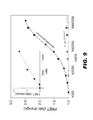
- FIG. 9 depicts a biosensor for Salmonella typhimurium .
- FRET signal produced by the 20 nM/25 nM mixture of antibodies labeled with fluorescein and Cy5-modified complementary signaling oligonucleotides at indicated amounts of cells (in 20 ⁇ l assay, 384-well microplate). Averages and standard deviations of 3 independent measurements are shown.
- FIG. 10 depicts the kinetics of a Salmonella typhimurium biosensor response. Indicated amounts of Salmonella cells were added to 20 nM/25 nM mixture of antibody labeled with fluorescein and Cy5-modified complementary oligonucleotides and FRET signal (emission at 670 nm with excitation at 488 nm) was monitored over time.
- FIG. 11 depicts the homogenous restriction-enzyme based signal amplification methodology compatible with homogenous nature of molecular biosensors.
- S 1 and S 2 correspond to the antibodies recognizing the target labeled with short oligonucleotides complementary to the region of S 3 flanking the restriction enzyme cleavage site in S 3 .
- FIG. 12 depicts a design of the solid-surface variant of the sensor for detecting bacteria. Oligonucleotide containing multiple copies of a sequence complementary to the signaling oligonucleotide used for labeling the antibody is immobilized on the solid surface.
- FIG. 13 depicts a proof-of-principle validation of the design illustrated in FIG. 12 .
- Various amounts of E. coli O157:H7 cells were incubated in the presence of 20 nM antibody conjugated to fluorescein-modified signaling oligonucleotide in microplate wells containing immobilized complementary oligonucleotide. After washing the wells with the buffer the fluorescence remaining in the wells were read on a plate reader.
- Inset Specificity of the solid-surface based sensor. No signal in the presence of E. coli K12 was detected (red symbols).
- FIG. 14 depicts a proof-of-principle for a TIRF-based solid-surface based biosensor for bacteria.
- Oligonucleotide containing multiple copies of a sequence complementary to the signaling oligonucleotide used for labeling the antibody was immobilized on the surface of a quartz slide.
- TIRF-induced fluorescence originating from the surface of the slide was monitored. Only a small signal was observed upon injecting the sample containing only labeled antibody whereas large signal was observed in the presence of target bacteria.
- FIG. 15 depicts two standard curves for a ferritin assay.
- the present invention is directed to a method of using a molecular biosensor.
- the method comprises detecting targets comprising repeating epitopes.
- the method typically involves target-molecule induced co-association of two epitope-binding agents that each recognize the same repeating epitope on the target.
- the epitope-binding agents each comprise complementary signaling oligonucleotides that are labeled with detection means and are attached to the epitope binding agents through a flexible linker. Co-association of the two epitope-binding agents with the target results in bringing the two signaling oligonucleotides into proximity such that a detectable signal is produced.
- the molecular biosensors provide a rapid homogeneous means to detect a variety of targets comprising repeating epitopes, including but not limited to proteins, carbohydrates, lipids, and microbial organisms.
- the molecular biosensor may be monovalent comprising a single epitope binding agent that binds to an epitope on a target.
- a molecular biosensor of the invention is typically multivalent. It will be appreciated by a skilled artisan, depending upon the target, that the molecular biosensor may comprise from about 2 to about 5 epitope binding agents. Typically, the molecular biosensor may comprise 2 or 3 epitope binding agents and more typically, may comprise 2 epitope binding agents.
- the molecular biosensor may be bivalent comprising a first epitope binding agent that binds to a repeating epitope on a target and a second epitope binding agent that binds to the same repeating epitope on the target.
- the molecular biosensor may be trivalent comprising a first epitope binding agent that binds to a repeating epitope on a target, a second epitope binding agent that binds to the same repeating epitope on a target and a third epitope binding agent that binds to the same repeating epitope on a target.
- the molecular biosensor may be bivalent.
- the bivalent construct will comprise a first epitope binding agent that binds to a repeating epitope on a target, a first linker, a first signaling oligo, a first detection means, a second epitope binding agent that binds to the same repeating epitope on the target, a second linker, a second signaling oligo, and a second detection means.
- the molecular biosensor comprises two nucleic acid constructs, which together have formula (I): R 1 —R 2 —R 3 —R 4 ; and R 5 —R 6 —R 7 —R 8 ; (I)
- epitope binding agents R 1 and R 5
- R 1 and R 5 in molecular biosensors having formula (I) can and will vary depending upon the particular target.
- R 1 and R 5 may be an aptamer, or antibody.
- R 1 and R 5 are double stranded nucleic acid the target is typically a macromolecule that binds to DNA or a DNA binding protein.
- suitable choices for R 1 and R 5 will include two agents that each recognize a repeating epitope on the same target. In certain embodiments, however, it is also envisioned that R 1 and R 5 may recognize repeating epitopes on different targets.
- Non-limiting examples of suitable epitope binding agents include agents selected from the group consisting of an aptamer, an antibody, an antibody fragment, a double-stranded DNA sequence, a ligand, a ligand fragment, a receptor, a receptor fragment, a polypeptide, a peptide, a coenzyme, a coregulator, an allosteric molecule, and an ion.
- R 1 and R 5 are each antibodies selected from the group consisting of polyclonal antibodies, ascites, Fab fragments, Fab′ fragments, monoclonal antibodies, chimeric antibodies, and humanized antibodies. In a preferred embodiment, R 1 and R 5 are each monoclonal antibodies.
- R 1 and R 5 are each aptamers. In an additional embodiment, R 1 and R 5 are each double stranded DNA. In a further embodiment, R 1 is a double stranded nucleic acid and R 5 is an aptamer. In an additional embodiment, R 1 is an antibody and R 5 is an aptamer. In an additional embodiment, R 1 is an antibody and R 5 is a double stranded DNA. In embodiments where R 1 and R 5 comprise the same epitope binding agent, the bivalent biosensor may be considered a monovalent biosensor.
- exemplary linkers, R 2 and R 6 will functionally keep R 3 and R 7 in close proximity such that when R 1 and R 5 each bind to the target, R 3 and R 7 associate in a manner such that a detectable signal is produced by the detection means, R 4 and R 8 .
- R 2 and R 6 may each be a nucleotide sequence from about 10 to about 100 nucleotides in length. In one embodiment, R 2 and R 6 are from 10 to about 25 nucleotides in length. In another embodiment, R 2 and R 6 are from about 25 to about 50 nucleotides in length. In a further embodiment, R 2 and R 6 are from about 50 to about 75 nucleotides in length.
- R 2 and R 6 are from about 75 to about 100 nucleotides in length.
- the nucleotides comprising the linkers may be any of the nucleotide bases in DNA or RNA (A, C, T, G in the case of DNA, or A, C, U, G in the case of RNA).
- R 2 and R 6 are comprised of DNA bases.
- R 2 and R 6 are comprised of RNA bases.
- R 2 and R 6 are comprised of modified nucleic acid bases, such as modified DNA bases or modified RNA bases.
- modified DNA or RNA bases examples include 2′-fluoro nucleotides, 2′-amino nucleotides, 5′-aminoallyl-2′-fluoro nucleotides and phosphorothioate nucleotides.
- R 2 and R 6 may be a polymer of bifunctional chemical linkers. In one embodiment the bifunctional chemical linker is heterobifunctional.
- Suitable heterobifunctional chemical linkers include sulfoSMCC (Sulfosuccinimidyl-4-(N-maleimidomethyl)cyclohexane-1-carboxylate), and lc-SPDP(N-Succinimidyl-6-(3′-(2-PyridylDithio)-Propionamido)-hexanoate).
- the bifunctional chemical linker is homobifunctional.
- Suitable homobifunctional linkers include disuccinimidyl suberate, disuccinimidyl glutarate, and disuccinimidyl tartrate. Additional suitable linkers are illustrated in the Examples, such as the phosphoramidate form of Spacer 18 comprised of polyethylene glycol.
- R 2 and R 6 are from 0 to about 500 angstroms in length. In another embodiment, R 2 and R 6 are from about 20 to about 400 angstroms in length. In yet another embodiment, R 2 and R 6 are from about 50 to about 250 angstroms in length.
- R 3 and R 7 are complementary nucleotide sequences having a length such that they preferably do not associate unless R 1 and R 5 bind to separate repeating epitopes on the target. When R 1 and R 5 bind to separate epitopes of the target, R 3 and R 7 are brought to relative proximity resulting in an increase in their local concentration, which drives the association of R 3 and R 7 .
- R 3 and R 7 may be from about 2 to about 20 nucleotides in length. In another embodiment, R 3 and R 7 are from about 4 to about 15 nucleotides in length. In an exemplary embodiment, R 3 and R 7 are from about 5 to about 7 nucleotides in length.
- R 3 and R 7 have a free energy for association from about 5.5 kcal/mole to about 8.0 kcal/mole as measured in the selection buffer conditions, defined below. In another embodiment, R 3 and R 7 have a free energy for association from about 6.0 kcal/mole to about 8.0 kcal/mole as measured in the selection buffer conditions defined below. In yet another embodiment, R 3 and R 7 have a free energy for association from about 7.0 kcal/mole to 8.0 kcal/mole in the selection buffer conditions. In a preferred embodiment, R 3 and R 7 have a free energy for association of 7.5 kcal/mole in the selection buffer conditions described below. Preferably, in each embodiment R 3 and R 7 are not complementary to R 1 and R 5 .
- R 4 and R 8 may together comprise several suitable detection means such that when R 3 and R 7 associate, a detectable signal is produced.
- Exemplary detections means suitable for use in the molecular biosensors may include FRET, fluorescence cross-correlation spectroscopy, flourescence quenching, fluorescence polarization, flow cytometry, scintillation proximity, luminescence resonance energy transfer, direct quenching, ground-state complex formation, chemiluminescence energy transfer, bioluminescence resonance energy transfer, excimer formation, colorimetric substrates detection, phosphorescence, electro-chemical changes, and redox potential changes.
- the molecular biosensor will have formula (I) wherein:
- Yet another embodiment of the invention encompasses a molecular biosensor having formula (I) wherein:
- a further embodiment of the invention encompasses a molecular biosensor having formula (I) wherein:
- the molecular biosensor may be trivalent.
- the trivalent construct may comprise a first epitope binding agent that binds to a repeating epitope on a target, a first linker, a first signaling oligo, a first detection means, a second epitope binding agent that binds to the same repeating epitope on the target, a second linker, a second signaling oligo, a second detection means, a third epitope binding agent that binds to the same repeating epitope on a target, a third linker, a third signaling oligo, and a third detection means.
- the molecular biosensor comprises three nucleic acid constructs, which together have formula (II): R 15 —R 14 —R 13 —R 9 —R 10 —R 11 —R 12 ; R 16 —R 17 —R 18 —R 19 ; and R 20 —R 21 —R 22 —R 23 (II)
- epitope binding agents R 9 , R 16 and R 20
- suitable choices for R 9 , R 16 and R 20 will include three agents that each recognize the same repeating epitope on the same target or on different targets.
- Non-limiting examples of suitable epitope binding agents include agents selected from the group consisting of an aptamer, an antibody, an antibody fragment, a double-stranded DNA sequence, a ligand, a ligand fragment, a receptor, a receptor fragment, a polypeptide, a peptide, a coenzyme, a coregulator, an allosteric molecule, and an ion.
- R 9 , R 16 and R 20 are each antibodies.
- exemplary linkers, R 10 and R 21 will functionally keep R 11 and R 22 in close proximity such that when R 9 and R 20 each bind to the repeating epitopes on the target(s), R 11 and R 22 associate in a manner such that a detectable signal is produced by the detection means, R 12 and R 23 .
- exemplary linkers, R 13 and R 17 will functionally keep R 14 and R 18 in close proximity such that when R 9 and R 16 each bind to the repeating epitopes on the target(s), R 14 and R 18 associate in a manner such that a detectable signal is produced by the detection means, R 15 and R 19 .
- the linkers utilized in molecular biosensors having formula (II) may each be a nucleotide sequence from about 10 to about 100 nucleotides in length. In one embodiment, the linkers are from 10 to about 25 nucleotides in length. In another embodiment, the linkers are from about 25 to about 50 nucleotides in length. In a further embodiment, the linkers are from about 50 to about 75 nucleotides in length. In yet another embodiment, the linkers are from about 75 to about 100 nucleotides in length.
- the nucleotides comprising the linkers may be any of the nucleotide bases in DNA or RNA (A, C, T, G in the case of DNA, or A, C, U, G in the case of RNA).
- the linkers are comprised of DNA bases.
- the linkers are comprised of RNA bases.
- the linkers are comprised of modified nucleic acid bases, such as modified DNA bases or modified RNA bases. Examples of suitable modified DNA or RNA bases include 2′-fluoro nucleotides, 2′-amino nucleotides, 5′-aminoallyl-2′-fluoro nucleotides and phosphorothioate nucleotides.
- the linkers may be a polymer of bifunctional chemical linkers.
- the bifunctional chemical linker is heterobifunctional.
- Suitable heterobifunctional chemical linkers include sulfoSMCC (Sulfosuccinimidyl-4-(N-maleimidomethyl)cyclohexane-1-carboxylate), and lc-SPDP(N-Succinimidyl-6-(3′-(2-PyridylDithio)-Propionamido)-hexanoate).
- the bifunctional chemical linker is homobifunctional.
- Suitable homobifunctional linkers include disuccinimidyl suberate, disuccinimidyl glutarate, and disuccinimidyl tartrate. Additional suitable linkers are illustrated in the Examples, such as the phosphoramidate form of Spacer 18 comprised of polyethylene glycol.
- the linkers are from 0 to about 500 angstroms in length. In another embodiment, the linkers are from about 20 to about 400 angstroms in length. In yet another embodiment, the linkers are from about 50 to about 250 angstroms in length.
- R 11 and R 22 are complementary nucleotide sequences having a length such that they preferably do not associate unless R 9 and R 20 bind to separate repeating epitopes on the target(s).
- R 14 and R 18 are complementary nucleotide sequences having a length such that they preferably do not associate unless R 9 and R 16 bind to separate repeating epitopes on the target(s).
- R 11 and R 22 and R 14 and R 18 may be from about 2 to about 20 nucleotides in length. In another embodiment, R 11 and R 22 and R 14 and R 18 are from about 4 to about 15 nucleotides in length.
- R 11 and R 22 and R 14 and R 18 are from about 5 to about 7 nucleotides in length. In one embodiment, R 11 and R 22 and R 14 and R 18 have a free energy for association from about 5.5 kcal/mole to about 8.0 kcal/mole as measured in the selection buffer conditions, defined below. In another embodiment, R 11 and R 22 and R 14 and R 18 have a free energy for association from about 6.0 kcal/mole to about 8.0 kcal/mole as measured in the selection buffer conditions defined below. In yet another embodiment, R 11 and R 22 and R 14 and R 18 have a free energy for association from about 7.0 kcal/mole to 8.0 kcal/mole in the selection buffer conditions.
- R 11 and R 22 and R 14 and R 18 have a free energy for association of 7.5 kcal/mole in the selection buffer conditions described below.
- R 11 and R 22 and R 14 and R 18 are not complementary to any of R 9 , R 16 or R 20 .
- R 12 and R 23 may together comprise several suitable detection means such that when R 11 and R 22 associate, a detectable signal is produced.
- R 15 and R 19 may together comprise several suitable detection means such that when R 14 and R 18 associate, a detectable signal is produced.
- Exemplary detections means suitable for use in the molecular biosensors include FRET, fluorescence cross-correlation spectroscopy, flourescence quenching, fluorescence polarization, flow cytometry, scintillation proximity, luminescence resonance energy transfer, direct quenching, ground-state complex formation, chemiluminescence energy transfer, bioluminescence resonance energy transfer, excimer formation, colorimetric substrates detection, phosphorescence, electro-chemical changes, and redox potential changes.
- Another embodiment of the invention comprises three-component molecular biosensors. These biosensors, in addition to having at least two epitope binding agent constructs, further comprise an oligonucleotide construct, referred to below as “O.” In certain embodiments, the three-component molecular biosensor will comprise an endonuclease restriction site. In alternative embodiments, the three-component molecular biosensor will not have an endonuclease restriction site.
- the three-component biosensor will comprise: (1) a first epitope binding agent construct that binds to a repeating epitope on a target, a first linker, a first signaling oligo, and a first detection means; (2) a second epitope binding agent construct that binds to the same repeating epitope on the target, a second linker, a second signaling oligo, and a second detection means; and (3) an oligonucleotide construct that comprises a first region that is complementary to the first oligo and a second region that is complementary to the second oligo.
- the first signaling oligo and second signaling oligo are not complementary to each other, but are complementary to two distinct regions on the oligonucleotide construct. Co-association of the two epitope-binding agent constructs with the target results in hybridization of each signaling oligos to the oligonucleotide construct. Binding of the two signaling oligo to the oligonucleotide construct brings them into proximity such that a detectable signal is produced.
- the three-component molecular biosensor comprises three nucleic acid constructs, which together have formula (III): R 24 —R 25 —R 26 —R 27 ; R 28 —R 29 —R 30 —R 31 ; O (III)
- epitope binding agents R 24 and R 28 in molecular biosensors having formula (III) can and will vary depending upon the particular target.
- R 24 and R 28 may be an aptamer, or antibody.
- R 24 and R 28 are double stranded nucleic acid the target is typically a macromolecule that binds to DNA or a DNA binding protein.
- suitable choices for R 24 and R 28 will include two agents that each recognize the same repeating epitope on the same target. In certain embodiments, however, it is also envisioned that R 24 and R 28 may recognize distinct epitopes on different targets.
- Non-limiting examples of suitable epitope binding agents include agents selected from the group consisting of an aptamer, an antibody, an antibody fragment, a double-stranded DNA sequence, modified nucleic acids, nucleic acid mimics, a ligand, a ligand fragment, a receptor, a receptor fragment, a polypeptide, a peptide, a coenzyme, a coregulator, an allosteric molecule, and an ion.
- R 24 and R 28 are each antibodies selected from the group consisting of polyclonal antibodies, ascites, Fab fragments, Fab′ fragments, monoclonal antibodies, chimeric antibodies, and humanized antibodies.
- R 24 and R 28 are each aptamers.
- R 24 and R 28 are peptides.
- R 24 and R 28 are each monoclonal antibodies.
- R 24 and R 28 are each double stranded DNA.
- R 24 is a double stranded nucleic acid and R 28 is an aptamer.
- R 24 is an antibody and R 28 is an aptamer.
- R 24 is an antibody and R 28 is a double stranded DNA.
- R 25 and R 29 may each be a nucleotide sequence from about 10 to about 100 nucleotides in length. In one embodiment, R 25 and R 29 are from 10 to about 25 nucleotides in length. In another embodiment, R 2 and R 6 are from about 25 to about 50 nucleotides in length. In a further embodiment, R 25 and R 29 are from about 50 to about 75 nucleotides in length. In yet another embodiment, R 25 and R 29 are from about 75 to about 100 nucleotides in length.
- the nucleotides comprising the linkers may be any of the nucleotide bases in DNA or RNA (A, C, T, G in the case of DNA, or A, C, U, G in the case of RNA).
- R 25 and R 29 are comprised of DNA bases.
- R 25 and R 29 are comprised of RNA bases.
- R 25 and R 29 are comprised of modified nucleic acid bases, such as modified DNA bases or modified RNA bases. Modifications may occur at, but are not restricted to, the sugar 2′ position, the C-5 position of pyrimidines, and the 8-position of purines.
- R 25 and R 29 may be nucleotide mimics.
- nucleotide mimics include locked nucleic acids (LNA), peptide nucleic acids (PNA), and phosphorodiamidate morpholino oligomers (PMO).
- R 25 and R 29 may be a polymer of bifunctional chemical linkers.
- the bifunctional chemical linker is heterobifunctional.
- Suitable heterobifunctional chemical linkers include sulfoSMCC (Sulfosuccinimidyl-4-(N-maleimidomethyl)cyclohexane-1-carboxylate), and lc-SPDP(N-Succinimidyl-6-(3′-(2-PyridylDithio)-Propionamido)-hexanoate).
- the bifunctional chemical linker is homobifunctional.
- Suitable homobifunctional linkers include disuccinimidyl suberate, disuccinimidyl glutarate, and disuccinimidyl tartrate. Additional suitable linkers are illustrated in the Examples, such as the phosphoramidate form of Spacer 18 comprised of polyethylene glycol.
- R 25 and R 29 are from 0 to about 500 angstroms in length. In another embodiment, R 25 and R 29 are from about 20 to about 400 angstroms in length. In yet another embodiment, R 25 and R 29 are from about 50 to about 250 angstroms in length.
- R 26 and R 30 are nucleotide sequences that are not complementary to each other, but that are complementary to two distinct regions of O.
- R 26 and R 30 may be from about 2 to about 20 nucleotides in length.
- R 26 and R 30 are from about 4 to about 15 nucleotides in length.
- R 26 and R 30 are from about 5 to about 7 nucleotides in length.
- R 26 and R 30 are not complementary to R 24 and R 28 .
- R 27 and R 31 may together comprise several suitable detection means such that when R 26 and R 30 each bind to complementary, distinct regions on O, a detectable signal is produced.
- Exemplary detections means suitable for use in the molecular biosensors include fluorescent resonance energy transfer (FRET), lanthamide resonance energy transfer (LRET), fluorescence cross-correlation spectroscopy, flourescence quenching, fluorescence polarization, flow cytometry, scintillation proximity, luminescence resonance energy transfer, direct quenching, ground-state complex formation, chemiluminescence energy transfer, bioluminescence resonance energy transfer, excimer formation, colorimetric substrates detection, phosphorescence, electro-chemical changes, and redox potential changes.
- FRET fluorescent resonance energy transfer
- LRET lanthamide resonance energy transfer
- fluorescence cross-correlation spectroscopy fluorescence cross-correlation spectroscopy
- flourescence quenching fluorescence polarization
- flow cytometry flow cytometry
- O comprises a first region that is complementary to R 26 , and a second region that is complementary to R 30 .
- O may be from about 8 to about 100 nucleotides in length.
- O is from about 10 to about 15 nucleotides in length, or from about 15 to about 20 nucleotides in length, or from about 20 to about 25 nucleotides in length, or from about 25 to about 30 nucleotides in length, or from about 30 to about 35 nucleotides in length, or from about 35 to about 40 nucleotides in length, or from about 40 to about 45 nucleotides in length, or from about 45 to about 50 nucleotides in length, or from about 50 to about 55 nucleotides in length, or from about 55 to about 60 nucleotides in length, or from about 60 to about 65 nucleotides in length, or from about 65 to about 70 nucleotides in length, or from about 70 to about 75 nucleotides in
- O will comprise formula (IV): R 32 —R 33 —R 34 —R 35 —R 36 (IV)
- R 33 and R 35 generally have a length such that the free energy of association between R 33 and R 26 and R 35 and R 30 is from about ⁇ 5 to about ⁇ 12 kcal/mole at a temperature from about 21° C. to about 40° C. and at a salt concentration from about 1 mM to about 100 mM.
- the free energy of association between R 33 and R 26 and R 35 and R 30 is about ⁇ 5 kcal/mole, about ⁇ 6 kcal/mole, about ⁇ 7 kcal/mole, about ⁇ 8 kcal/mole, about ⁇ 9 kcal/mole, about ⁇ 10 kcal/mole, about ⁇ 11 kcal/mole, or greater than about ⁇ 12 kcal/mole at a temperature from about 21° C. to about 40° C. and at a salt concentration from about 1 mM to about 100 mM.
- R 33 and R 35 may range from about 4 to about 20 nucleotides in length. In other embodiments, R 33 and R 35 may about 4, about 5, about 6, about 7, about 8, about 9, about 10, about 11, about 12, about 13, about 14, about 15, about 16, about 17, about 18, about 19, or greater than about 10 nucleotides in length.
- the three-component biosensor will comprise: (1) a first epitope binding agent construct that binds to a repeating epitope on a target, a first linker, and a first signaling oligo; (2) a second epitope binding agent construct that binds to the same repeating epitope on the target, a second linker, a second signaling oligo and (3) an oligonucleotide construct that comprises a first region that is complementary to the first oligo, a second region that is complementary to the second oligo, two flexible linkers, an endonuclease restriction site overlapping the first and the second regions complementary to the first and the second oligos, and a pair of complementary nucleotides with detection means.
- the first signaling oligo and second signaling oligo are not complementary to each other, but are complementary to two distinct regions on the oligonucleotide construct.
- the complementary nucleotides are annealed and produce a detectable signal. Co-association of the two epitope-binding agent constructs with the target results in hybridization of each signaling oligo to the oligonucleotide construct.
- the signaling oligos hybridize to two distinct locations on the oligonucleotide construct such that a double-stranded DNA molecule containing the restriction site is produced, with a gap between the signaling oligos located exactly at the site of endonuclease cleavage in one strand of the double-stranded DNA substrate.
- a restriction endonuclease When a restriction endonuclease is present, accordingly, it will cleave the oligonucleotide construct only when the target is present (i.e., when the signaling oligos are bound to the oligonucleotide construct). Upon this cleavage, the detection means present on the oligonucleotide are separated-resulting in no detectable signal.
- another oligonucleotide construct may hybridize with the signaling oligos of the two epitope-binding agents co-associated with the target and the cleavage reaction may be repeated. This cycle of hybridization and cleavage may be repeated many times resulting in cleavage of multiple oligonucleotide constructs per one complex of the two epitope-binding agents with the target.
- the three-component molecular biosensor comprises three nucleic acid constructs, which together have formula (V): R 36 —R 37 —R 38 ; R 39 —R 40 —R 41 ; O (V)
- Suitable linkers, epitope binding agents, and detection means for three-component molecular biosensors having formula (V) are the same as three component molecular biosensors having formula (III).
- Suitable, endonuclease restriction sites comprising R 42 include sites that are recognized by restriction enzymes that cleave double stranded nucleic acid, but not single stranded nucleic acid.
- these sites may include Accl, Agel, BamHI, Bgl, BglI, BsiWI, BstBI, ClaI, CviQI, Ddel, DpnI, DraI, EagI, EcoRI, EcoRV, FseI, FspI, HaeII, HaeIII, HhaI, Hinc II, HinDIII, HpaI, HpaII, KpnI, KspI, MboI, MfeI, NaeI, NarI, NcoI, NdeI, NheI, NotI, PhoI, PstI, PvuI, PvuII, SacI, SadI, SalI, SbfI, SmaI, SpeI, SphI, StuI, TaqI, TfiI, TliI, XbaI, XhoI, XmaI, XmnI, and ZraI.
- R 42 may comprise nucleotide spacers that precede or follow one or more of the endonuclease restriction site, the first region that is complementary to R 38 , and/or the second region that is complementary to R 41 .
- Suitable nucleotide spacers for example, are detailed in formula (IV).
- a further aspect of the invention encompasses the use of the molecular biosensors of the invention in several applications.
- the molecular biosensors are utilized in methods for detecting one or more targets.
- the molecular biosensors may be utilized in kits and for therapeutic applications.
- a signal produced by a biosensor of the invention may be detectable in about 1, 2, 3, 4, 5, 6, 7, 8, 9, or 10 minutes. In certain embodiments, 80% of the maximum signal may be produced in about 3, 4, 5, 6, 7 or 8 minutes. In other embodiments, the maximum signal may be produced in about 5, 10, 15, 20, 25, 30, 35, 40, or 45 minutes.
- the molecular biosensors may be utilized for the detection of a target.
- the method generally involves contacting a molecular biosensor of the invention with the target.
- the method typically involves target-molecule induced co-association of two epitope-binding agents (present on the molecular biosensor of the invention) that each recognize the same repeating epitope on the target.
- the epitope-binding agents each comprise complementary signaling oligonucleotides that are labeled with detection means and are attached to the epitope binding agents through a flexible linker.
- Co-association of the two epitope-binding agents with the target results in bringing the two signaling oligonucleotides into proximity such that a detectable signal is produced.
- the detectable signal is produced by any of the detection means known in the art or as described herein.
- a method for the detection of a target that is a protein or polypeptide comprises a repeating epitope.
- the method generally involves detecting a protein or polypeptide in a sample comprising the steps of contacting a sample with a molecular biosensor of the invention.
- the molecular biosensors may be used to detect a target that is a macromolecular complex in a sample.
- the repeating epitope is preferably on each component of the macromolecular complex, such that when the complex is formed, the epitope-binding agents that recognize the repeating epitope are bought into proximity, resulting in the stable interaction of the first signaling oligo and the second signaling oligo to produce a detectable signal, as described above.
- the invention also encompasses a solid surface having one or more of the molecular constructs of the invention attached thereto.
- suitable surfaces include microtitre plates, test tubes, beads, resins and other polymers, as well as other surfaces either known in the art or described herein.
- the solid surface comprises a nucleic acid sequence that is complementary to a signaling oligo of one or more molecular constructs, as described in the examples. Therefore, when the epitope binding agent recognizes the target comprising repeating epitopes, the signaling oligo of the molecular construct binds to the immobilized nucleic acid sequence. The binding may be detected by any means detailed above or in the examples.
- the solid surface utilizes a three-component biosensor.
- the oligonucleotide construct may be immobilized on a solid surface.
- the first epitope binding agent and second epitope binding agent are contacted with the surface comprising immobilized O and a sample that may comprise a target.
- the first epitope binding agent, second epitope binding agent, and target bind to immobilized O to form a complex.
- Several methods may be utilized to detect the presence of the complex comprising target. The method may include detecting a probe attached to the epitope-binding agents after washing out the unbound components.
- several surface specific real-time detection methods may be employed, including but not limited to surface plasmon resonance (SPR) or total internal reflection fluorescence (TIRF).
- the oligonucleotide construct, O may be immobilized to several types of suitable surfaces.
- the surface may be a material that may be modified to contain discrete individual sites appropriate for the attachment or association of the three-component biosensor and is amenable to at least one detection method.
- suitable surfaces include glass, modified or functionalized glass, plastics (including acrylics, polystyrene and copolymers of styrene and other materials, polypropylene, polyethylene, polybutylene, polyurethanes, TeflonJ, etc.), nylon or nitrocellulose, polysaccharides, nylon, resins, silica or silica-based materials including silicon and modified silicon, carbon, metals, inorganic glasses and plastics.
- a surface may be planar, a surface may be a well, i.e. a 364 well plate, or alternatively, a surface may be a bead or a slide.
- the oligonucleotide construct, O may be attached to the surface in a wide variety of ways, as will be appreciated by those in the art.
- O for example, may either be synthesized first, with subsequent attachment to the surface, or may be directly synthesized on the surface.
- the surface and O may be derivatized with chemical functional groups for subsequent attachment of the two.
- the surface may be derivatized with a chemical functional group including, but not limited to, amino groups, carboxyl groups, oxo groups or thiol groups. Using these functional groups, the O may be attached using functional groups either directly or indirectly using linkers. Alternatively, O may also be attached to the surface non-covalently.
- a biotinylated O can be prepared, which may bind to surfaces covalently coated with streptavidin, resulting in attachment.
- O may be synthesized on the surface using techniques such as photopolymerization and photolithography. Additional methods of attaching O to a surface and methods of synthesizing O on surfaces are well known in the art, i.e. VLSIPS technology from Affymetrix (e.g., see U.S. Pat. No. 6,566,495, and Rockett and Dix, “DNA arrays: technology, options and toxicological applications,” Xenobiotica 30(2):155-177, all of which are hereby incorporated by reference in their entirety).
- the molecular biosensor may not include a detections means.
- the molecular biosensor when the molecular biosensor is a bivalent antibody construct, the bivalent antibody construct may not have labels for detection. It is envisioned that these alternative bivalent constructs may be used much like antibodies to detect molecules, bind molecules, purify molecules (as in a column or pull-down type of procedure), block molecular interactions, facilitate or stabilize molecular interactions, or confer passive immunity to an organism. It is further envisioned that the bivalent antibody construct can be used for therapeutic purposes.
- This invention enables the skilled artisan to build several combinations of antibodies that recognize any repeating epitope from any number of molecules into a bivalent, trivalent, or other multivalent aptamer construct to pull together those disparate molecules to test the effect or to produce a desired therapeutic outcome.
- the invention is directed to a kit comprising a first epitope binding agent, to which is attached a first label, and a second epitope binding agent, to which is attached a second label, wherein (a) when the first epitope binding agent and the second epitope binding agent bind to a repeating epitope of the polypeptide, (b) the first label and the second label interact to produce a detectable signal.
- the epitope-binding agent is an antibody construct, which comprises an antibody, a label and a signaling oligo.
- the epitope-binding agent may be an antibody fragment, an aptamer, or a peptide.
- the kit is useful in the detection of targets comprising a repeating epitope, and as such, may be used in research or medical/veterinary diagnostics applications.
- the kit may be used to detect microbial organisms, such as bacteria, viruses, and fungi.
- the invention is directed to a method of diagnosing a disease comprising the steps of (a) obtaining a sample from a patient, (b) contacting the sample with a first epitope binding agent construct and a second epitope binding agent construct, and (c) detecting the presence of a polypeptide, microbial organism, or macromolecular complex in the sample using a detection method, wherein the presence of the polypeptide, microbial organism, or macromolecular complex in the sample indicates whether a disease is present in the patient.
- the first epitope binding agent construct is a first antibody to which a first label and a first signaling oligo are attached
- the second epitope binding agent construct is a second antibody to which a second label and a second signaling oligo, which is complementary to the first signaling oligo, are attached
- the detection method is a fluorescence detection method, wherein, (d) when the first antibody binds to the polypeptide and the second antibody binds to the polypeptide, (e) the first signaling oligo and the second signaling oligo associate with each other, and (f) the first label is brought into proximity to the second label such that a change in fluorescence occurs.
- the first epitope binding agent construct is a first peptide to which a first label and a first signaling oligo are attached
- the second epitope binding agent construct is a second peptide to which a second label and a second signaling oligo, which is complementary to the first signaling oligo
- the detection method is a fluorescence detection method, wherein, (d) when the first peptide binds to the polypeptide and the second peptide binds to the polypeptide, (e) the first signaling oligo and the second signaling oligo associate with each other, and (f) the first label is brought into proximity to the second label such that a change in fluorescence occurs.
- the first epitope binding agent and the second epitope-binding agents are different types of epitope binding agents (i.e. an antibody and a peptide, an aptamer and an antibody, etc.).
- Preferred samples include blood, urine, ascites, cells and tissue samples/biopsies.
- Preferred patients include humans, farm animals and companion animals.
- the invention is directed to a method of screening a sample for targets comprising the steps of (a) contacting a sample with a first epitope binding agent construct and a second epitope binding agent construct, and (b) detecting the presence of a target in the sample using a detection method.
- Preferred targets include a polypeptide, which comprises a repeating epitope, and a microbial organism.
- the first epitope binding agent is a first antibody to which a first label and a first signaling oligo are attached
- the second epitope binding agent is a second antibody to which a second label and a second signaling oligo, which is complementary to the first signaling oligo, are attached
- the detection method is a fluorescence detection method, wherein, (d) when the first antibody binds to the polypeptide and the second antibody binds to the polypeptide, (e) the first signaling oligo and the second signaling oligo associate with each other, and (f) the first label is brought into proximity to the second label such that a change in fluorescence occurs.
- the first epitope binding agent is a first peptide to which a first label and a first signaling oligo are attached
- the second epitope binding agent is a second peptide to which a second label and a second signaling oligo, which is complementary to the first signaling oligo
- the detection method is a fluorescence detection method, wherein, (d) when the first peptide binds to the polypeptide and the second peptide binds to the polypeptide, (e) the first signaling oligo and the second signaling oligo associate with each other, and (f) the first label is brought into proximity to the second label such that a change in fluorescence occurs.
- the first epitope binding agent and the second epitope-binding agents are different types of epitope binding agents (i.e. an antibody and a peptide, an aptamer and an antibody, etc.).
- antibody generally means a polypeptide or protein that recognizes and can bind to an epitope of an antigen.
- An antibody as used herein, may be a complete antibody as understood in the art, i.e., consisting of two heavy chains and two light chains, or be selected from a group comprising polyclonal antibodies, ascites, Fab fragments, Fab′ fragments, monoclonal antibodies, humanized antibodies, and a peptide comprising a hypervariable region of an antibody.
- aptamer refers to a polynucleotide, generally a RNA or a DNA that has a useful biological activity in terms of biochemical activity, molecular recognition or binding attributes. Usually, an aptamer has a molecular activity such as binding to a target at a specific epitope (region). It is generally accepted that an aptamer, which is specific in its binding to any polypeptide, may be synthesized and/or identified by in vitro evolution methods.
- detection method means any of several methods known in the art to detect a molecular interaction event.
- the phrase “detectable signal”, as used herein, is essentially equivalent to “detection method.”
- Detection methods include detecting changes in mass (e.g., plasmin resonance), changes in fluorescence (e.g., FRET, FCCS, fluorescence quenching or increasing fluorescence, fluorescence polarization, flow cytometry), enzymatic activity (e.g., depletion of substrate or formation of a product, such as a detectable dye—NBT-BCIP system of alkaline phosphatase is an example), changes in chemiluminescence or scintillation (e.g., scintillation proximity assay, luminescence resonance energy transfer, bioluminescence resonance energy transfer and the like), and ground-state complex formation, excimer formation, colorimetric substance detection, phosphorescence, electro-chemical changes, and redox potential changes.
- mass e.g.,
- epitope refers generally to a particular region of a target. Examples include an antigen, a hapten, a molecule, a polymer, a prion, a microbe, a cell, a peptide, polypeptide, protein, a ligand, a receptor, or macromolecular complex.
- An epitope may consist of a small peptide derived from a larger polypeptide.
- An epitope may be a two or three-dimensional surface or surface feature of a polypeptide, protein or macromolecular complex that comprises several non-contiguous peptide stretches or amino acid groups.
- epitope binding agent refers to a substance that is capable of binding to a specific epitope of an antigen, a polypeptide, a protein or a macromolecular complex.
- epitope binding agents may include aptamers, double-stranded DNA sequence, ligands and fragments of ligands, receptors and fragments of receptors, antibodies and fragments of antibodies, polynucleotides, coenzymes, coregulators, allosteric molecules, and ions.
- epitope binding agent construct refers to a construct that contains an epitope-binding agent and can serve in a “molecular biosensor” with another molecular biosensor.
- an epitope binding agent construct also contains a “linker,” and a “signaling oligo”.
- Epitope binding agent constructs can be used to initiate the aptamer selection methods of the invention.
- a first epitope binding agent construct and a second epitope binding agent construct may be joined together by a “linker” to form a “bivalent epitope binding agent construct.”
- An epitope binding agent construct can also be referred to as a molecular recognition construct.
- An aptamer construct is a special kind of epitope binding agent construct wherein the epitope binding agent is an aptamer.
- label refers to any substance attachable to a polynucleotide, polypeptide, aptamer, nucleic acid component, or other substrate material, in which the substance is detectable by a detection method.
- Non-limiting examples of labels applicable to this invention include but are not limited to luminescent molecules, chemiluminescent molecules, fluorochromes, fluorescent quenching agents, colored molecules, radioisotopes, scintillants, massive labels (for detection via mass changes), biotin, avidin, streptavidin, protein A, protein G, antibodies or fragments thereof, Grb2, polyhistidine, Ni2+, Flag tags, myc tags, heavy metals, enzymes, alkaline phosphatase, peroxidase, luciferase, electron donors/acceptors, acridinium esters, and colorimetric substrates.
- the skilled artisan would readily recognize other useful labels that are not mentioned above, which may be employed in the operation of the present invention.
- Macromolecular complex refers to a composition of matter comprising a macromolecule.
- these are complexes of one or more macromolecules, such as polypeptides, lipids, carbohydrates, nucleic acids, natural or artificial polymers and the like, in association with each other. The association may involve covalent or non-covalent interactions between components of the macromolecular complex.
- Macromolecular complexes may be relatively simple, such as a ligand bound polypeptide, relatively complex, such as a lipid raft, or very complex, such as a cell surface, virus, bacteria, spore and the like.
- Macromolecular complexes may be biological or non-biological in nature.
- molecular biosensor and “molecular beacon” are used interchangeably herein to refer to a construct comprised of at least two epitope binding agent constructs.
- the molecular biosensor can be used for detecting or quantifying the presence of a target using a chemical-based system for detecting or quantifying the presence of an analyte, a prion, a protein, a nucleic acid, a lipid, a carbohydrate, a biomolecule, a macromolecular complex, a fungus, a microbial organism, or a macromolecular complex comprised of biomolecules using a measurable read-out system as the detection method.
- signaling oligo means a short (generally 2 to 15 nucleotides, preferably 5 to 7 nucleotides in length) single-stranded polynucleotide.
- Signaling oligos are typically used in pairs comprising a first signaling oligo and a second signaling oligo.
- the first signaling oligo sequence is complementary to the second signaling oligo.
- the first signaling oligo and the second signaling oligo can not form a stable association with each other through hydrogen bonding unless the first and second signaling oligos are brought into close proximity to each other through the mediation of a third party agent.
- FIG. 1 The overall design of molecular biosensors is illustrated in FIG. 1 .
- This concept is derived from molecular beacons for detecting DNA binding proteins and from aptamer-based molecular beacons for detecting proteins that were previously developed [12-15].
- a pair of antibodies recognizing non-overlapping epitopes of the protein is labeled with a pair of short complementary signaling oligonucleotides using a long flexible PEG-based crosslinker, respectively.
- Oligonucleotides are modified with a pair of fluorophores that may function as a donor and an acceptor in Fluorescence Resonance Energy Transfer (FRET) [16].
- FRET Fluorescence Resonance Energy Transfer
- Cardiac troponin I has been used as a target protein to demonstrate feasibility of the sensors depicted in FIG. 1A .
- the labeled antibodies were mixed in the absence of cardiac troponin, no significant FRET signal was observed when fluorescence spectra of donor-labeled antibody in the absence and the presence of acceptor-labeled antibody were compared ( FIG. 1B ).
- a large FRET signal was observed illustrated by several fold increase of emission at 670 nm with the excitation at 490 nm and quenching of donor emission at 520 nm. This signal allowed very sensitive (down to ⁇ 40 pM troponin) detection of the protein ( FIG. 1C ). Specificity of the sensor for cardiac troponin was tested.
- FIG. 2 By analogy to the target-protein induced annealing of signaling oligonucleotides ( FIG. 1 ), it was hypothesized that a similar effect could be produced by binding of signaling oligonucleotide-labeled antibodies to the repeating epitopes on a surface of a bacterial cell ( FIG. 2 ). Taking into account the dimensions of the bacterial cell ( ⁇ 500- ⁇ 3000 nm) and the length of flexible linkers used to attach the oligonucleotides to the antibodies (10-50 nm), it was expected that even a moderate density of cell surface epitopes recognized by the antibody should be sufficient to bring the antibodies into the proximity that would allow annealing of the signaling oligonucleotides.
- Complementary oligonucleotides provide additional favorable binding energy when the antibodies labeled with complementary oligonucleotides bind next to each other. This energy is absent when the two neighboring antibodies are labeled with the same signaling oligonucleotide (an arrangement that does not generate FRET signal).
- the design has a built-in selection process whereby the antibodies capable of producing FRET signal will be preferentially bound near each other. The preliminary data described below has fully validated the design illustrated in FIG. 2
- FIG. 4 shows the results of the three independent repeats of the assay performed at a wide range of target cells amounts. Few thousands of target cells were easily detectable using these assay conditions with high reproducibility (mean CV was 1.7%; range: 0.7-3.9%). No signal with negative control ( E. coli K12) has been detected at any amount of the cells tested.
- the FRET signal started to decrease most likely due to the decrease in the average amount of antibodies bound to each cell due to the depletion of the free antibody in the reaction mixture.
- Data shown in FIG. 3A and FIG. 4 demonstrate the feasibility of the assay design illustrated in FIG. 2 .
- the assay design illustrated in FIG. 2 assumes that FRET signal is generated by annealing of complementary oligonucleotides attached to the antibodies co-binding to the same cell.
- the other possibility could be that the FRET signal could be produced by annealing of signaling oligonucleotides attached to the antibodies bound to different cells (i.e. resulting in antibody-induced aggregation of the cells).
- Detection sensitivity will be one of the important parameters that will define the practical utility of the biosensors.
- One way to optimize assay sensitivity could be to find optimal concentrations of the antibodies. Reducing antibody concentrations could increase assay sensitivity since a bigger fraction of the total antibody concentration may be bound to cells at a given target cell density (provided that the antibody concentrations are not reduced below the Kd of antibody-antigen complex).
- FIG. 7 shows the response of the biosensor at two (lower) antibody concentrations. As expected, lowering antibody concentrations allowed detection of lower target cell amounts. It is estimated that using a 5/6.2 nM antibody mixture the sensitivity of detection was ⁇ 150 cells/20 ml assay.
- Methods for bacteria detection often employ some form of sample enrichment step to enhance detection of low cell amounts.
- a simple test was performed to demonstrate that such step can be added to the assay.
- Two diluted samples of E. coli O157:H7 were prepared.
- One (3000 cells/ml; 60 cells/20 ml assay) was below the detection limit using a sensor employing 20/25 nM mix of labeled antibodies ( FIG. 8 ).
- the other (300,000 cells/ml; 6000 cells/20 ml assay) gave relatively low signal with 20/25 nM mix of labeled antibodies ( FIG. 8 ).
- Both samples were then concentrated ⁇ 10 fold by a quick spin on a spin column with a semipermeable membrane and were re-measured. After the spin column treatment, the sample that was previously undetectable produced easy to measure signal, whereas the signal for the more concentrated sample (that previously gave relatively low reading) was dramatically improved ( FIG. 8 ).
- FIG. 9 shows that as in the case of E. coli O157:H7, a large dose-dependent FRET signal was observed with Salmonella cells. In contrast, very little FRET signal in the presence of E. coli K12 or E. coli O157:H7 was observed demonstrating the specificity of the sensor for the target cells. As in the case of E. coli O157:H7 reproducibility was excellent (mean CV was 1.0%; range: 0.3-2.2%). FRET signal in the case of Salmonella was smaller compared to E.
- Preparation of the antibodies modified with short oligonucleotides via long flexible linkers is an important step in preparing the biosensors for bacteria.
- a reliable procedure to label and purify the antibodies has been developed. Briefly, the antibody is treated with Traut's reagent to introduce free —SH groups. 5′-amino labeled oligonucleotide is reacted with NHS-maleimide bifunctional crosslinker producing maleimide-containing oligonucleotide that is reacted with —SH containing antibody.
- a long flexible linker is introduced to the oligonucleotide either during standard oligonucleotide synthesis [12] or can be a part of the bifunctional crosslinker. Labeled antibody is purified by FPLC size exclusion chromatography.
- Evaluating the pros and cons of various sensor design variants will involve performing the assays with a series of samples containing a range of dilution of each of the bacterial targets. Performance of the sensor will be evaluated by determining signal-to-background ratios, sensitivity and specificity of the detection. Sensitivity will be defined as the lowest amount of the target producing a signal higher than 3 ⁇ standard deviation over the background measured with no target. Specificity will be evaluated by comparing the signals measured in the presence of a given target with the signal measured at the same amount of unrelated bacteria.
- FIG. 2 schematically depicts antibodies labeled with one oligonucleotide per protein molecule. In reality, this does not need to be the case—it is unnecessary to prepare a homogenous preparation of such labeled antibodies.
- Degree of labeling of the antibodies can be modulated by changing the concentrations of the reagents and/or the time of coupling reactions.
- the performance of the sensors with antibodies labeled with oligonucleotides to a different degree can be compared to find the optimal ratio.
- the length of the oligonucleotides used to label the antibody will be important for optimal functioning of the sensors. To be more precise, it is not the length but the free energy change for their hybridization to the complementary strand that is the parameter of interest. If the binding affinity between the two complementary oligonucleotides used to label the antibody is too large, they will exhibit excessive target-independent association resulting in high background signal. If the affinity is too small, the background binding will be small but the target-induced annealing of the oligonucleotides will be reduced resulting in suboptimal FRET signal in the presence of target cells. In preliminary experiments, oligonucleotides with hybridization ⁇ G at 0.1 M NaCl of ⁇ 7.5 kcal/mole were used.
- oligonucleotide-labeled antibodies have been stored frozen at ⁇ 80°. When stored at 4°, they retain their functionality for at least 2 weeks. These observations demonstrate that the stability of the main component of the sensors is similar to any other immunological assay and is thus compatible with practical applications of the sensors.
- Homogenous assays in microplates can be greatly simplified by applying a dry-chemistry approach [17, 18]. In this approach the assay mixture solution is dried into the wells of the microplate. If the assay reagents are compatible with this procedure, the assay is simplified since it requires only a simple addition of the sample solution to a microplate well with the dried assay components. The shelf life of the assay can be increased and the storage requirements simplified when dry-chemistry approach is applied.
- the performance of the sensors will be tested using a dry-chemistry approach.
- the sensor mixtures (5 ml labeled antibody solution in a buffer containing 5% sorbitol which was shown to facilitate retention of functional properties of assay reagents after drying [18]) will be evaporated in the wells of 384-well microplate overnight in a desiccator over silica gel [17, 18].
- the sensitivity of the assays for the four bacterial targets will be compared using the dry-chemistry assay with the assays performed using the “normal” wet chemistry approach. If no significant decrease of assay sensitivity due to drying and dissolution of the assay components are observed, the stability of the assay in dry-chemistry format will be further tested.
- the plates with dry assay components sealed with a plate sealing film will be stored at room temperature or at 4° for an extended period of time (24 weeks).
- the performance of the stored dry-chemistry assays will be evaluated in weekly intervals to determine the effect of time of storage and storage temperature on the assay performance.
- Sensitivity of target cell detection will define where and how the sensors can be ultimately used.
- Current sensitivity of the sensors as determined in the preliminary experiments is equal or better to ELISA assays for bacteria detection [2].
- ELISA assays for bacteria detection [2].
- the sample potentially could be enriched using semi-permeable membrane concentration step (as illustrated by preliminary data illustrated by FIG. 8 ) or an cell culture enrichment step could be performed. Nevertheless, the more sensitive the sensors will be, the more applications they could potentially find.
- the following ideas for improving assay sensitivity may thus be tested:
- Time-resolved FRET (ii) Time-resolved FRET (LRET).
- LRET Luminescence Energy Transfer
- LRET is also very effective in removing the background due to light scattering or due to fluorescent impurities in the sample. This could be beneficial when turbid or autofluorescent samples need to be analyzed.
- antibodies labeled with complementary oligonucleotides modified with europium chelate and with Cy5, respectively, will be prepared. The performance of the sensors utilizing LRET-based detection will be compared with the sensors utilizing standard FRET detection.
- Hinc II sequence will be used but any restriction enzyme that cleaves ds DNA but is inactive on ss DNA (like Hinc II) could be used.
- S 3 also contains the probes attached to two complementary oligonucleotides that are in turn attached to S 3 via flexible linkers. When S 3 oligonucleotide is intact, the complementary oligonucleotides will be annealed (generating proximity-dependent signal such as, for example, FRET) due to the high local concentration resulting from their attachment to S 3 . Oligonucleotides used in S 1 and S 2 are designed to be sufficiently short that on their own they cannot anneal to S 3 in any significant manner.
- S 1 and S 2 in a complex with the target will bind to S 3 because the complex becomes effectively a bivalent binder for S 3 that will have much increased S 3 binding affinity compared to free S 1 or S 2 .
- the oligonucleotides of S 1 and S 2 are designed to anneal to S 3 such that a gap between the two oligonucleotides hybridized to S 3 is exactly at the position where Hinc II would normally cleave the bottom strand of DNA duplex. Thus, when Hinc II is present in the sample, it will cleave S 3 only when it is annealed to both S 1 and S 2 (i.e. when the target is present).
- the complex Upon cleavage of S 3 , the complex will dissociate (cleavage of S 3 will greatly decrease both the stability of the complex as well as it will result in dissociation of the two signaling oligonucleotides which in turn will eliminate the proximity-dependent signal).
- the complex of T with S 1 and S 2 can now associate with another molecule of S 3 and such cleavage and dissociation cycle could be repeated many times producing signal amplification.
- the same events as those illustrated in FIG. 11 for a protein target in solution could be produced by the antibodies associating with the surface of the bacteria cell (as illustrated in FIG. 2 ).
- the goal of this example will be to develop an alternative design of the sensor illustrated in FIG. 12 . While implementing this sensor design will require a more sophisticated instrumentation, its potential advantages include an increase in sensitivity and the possibility for multiplexed simultaneous detection of several targets.
- an antibody modified with a single short oligonucleotide via a long flexible linker will be used ( FIG. 12 ).
- the second oligonucleotide containing a segment(s) complementary to this oligonucleotide will be immobilized on a solid surface.
- the oligonucleotide that will be used to label the antibody will be designed to provide some affinity for binding to the immobilized complementary oligonucleotide but this affinity will be set (affinity of the interaction between two oligonucleotides can be set at any desired level using length and the sequence of oligonucleotides as a variable) to be low enough that at nanomolar concentrations of the antibody very little association of the free antibodies with the immobilized oligonucleotide will be observed (for example, a KD in mM range would fulfill this design requirement).
- FIG. 12 illustrates a feasibility experiment demonstrating detection of target bacteria with the solid-surface assay according to a design illustrated in FIG. 12 .
- oligonucleotide complementary to the antibody-bound oligonucleotide will be immobilized in the well of a 96-well microplate.
- the sample will be incubated with oligonucleotide-labeled antibody followed by washing the wells with the buffer and readout of fluorescence remaining in the well.
- Second variant will involve a real-time readout of the association of the target cell-antibody complex with the solid surface using Total Internal Reflection Fluorescence (TIRF).
- TIRF Total Internal Reflection Fluorescence
- This mode of signal readout will involve more technically complex instrumentation but will likely be more sensitive and faster. Additionally, it would be possible to immobilize oligonucleotides of unique sequences at different areas (spots) of TIRF slide. This would allow a simultaneous multiplexed detection of several target cells by using antibodies specific to each of the target cell labeled with a unique sequence oligonucleotide complementary to oligonucleotide immobilized at a unique spot of the slide.
- the optimal length of the oligonucleotides attached to the antibody in the case of the assay illustrated in FIG. 12 is likely to be different than in the homogenous assay ( FIG. 2 ). It is likely that lower affinity oligonucleotides may be required for optimal performance in the case of solid-surface based assay.
- the performance of the solid-surface variant of the assay ( FIG. 12 ) will therefore be tested utilizing the oligonucleotides with hybridization ⁇ G's in the range from 5 to 8 kcal/mole.
- oligonucleotides complementary to the antibody-bound oligonucleotides will be immobilized in the well of a 96-well microplate.
- This version of the solid-surface based assay is relatively easy to set up and perform and the conclusions regarding the optimal length of the complementary oligonucleotides obtained with this assay variant should be also applicable to the real-time TIRF-based assay. Streptavidin-coated plates and biotinylated oligonucleotide will be used to immobilize the oligonucleotides on a surface of the microplate well.
- oligonucleotide length tested various amounts of target cells and in parallel, negative control cells, will be incubated in the wells of the microplate coated with the corresponding complementary oligonucleotide. The wells will be washed three times with the buffer and fluorescence remaining in each well will be measured on Analyst AD microplate reader (Molecular Devices, Sunnyvale, Calif.). Sensitivity of detection and signal-to-background ratio for each oligonucleotide length tested will be used to determine the optimal length of the oligonucleotide.
- Immobilized oligonucleotides containing multiple repeats of the sequence complementary to the oligonucleotide used to label the antibody will also be tested (as opposed to a single copy of such sequence as depicted in FIG. 12 ).
- the affinity of the target cell coated with oligonucleotide-labeled antibodies for binding to the solid-surface containing oligonucleotides with multiple repeats of the complementary sequence may be greatly improved which would be beneficial for the performance of solid-surface based assay.
- TIRF Total Internal Reflection Fluorescence
- TIRF can be thus adopted for real-time detection of target cell induced association of the antibody-coated cell to the slide surface coated with the oligonucleotides complementary to the oligonucleotide used to label the antibody.
- Real-time detection capability will allow rapid readout time and the outstanding signal detection sensitivity of TIRF can produce significant sensitivity gains.
- FIG. 14 illustrates the feasibility of experiments demonstrating detection of target bacteria with the solid-surface assay implementing TIRF detection.
- TIRF-based detection offers a unique potential of developing a multiplexed assay that will allow simultaneous detection of multiple target cells.
- four oligonucleotides of unique sequence will be immobilized to four distinct areas (“spots”) on the TIRF slide.
- the antibodies for the four targets will be labeled, each with a unique sequence oligonucleotide complementary to the oligonucleotide immobilized in one of the spots.
- virus particles while much smaller then bacterial cells (for example, influenza virus is 80-120 nm in diameter), should be still large enough to produce FRET signal resulting from co-binding of many labeled antibodies to surface epitopes of the virus according to scheme in FIG. 2 .
- Influenza A and B viruses will be used as model targets to investigate the applicability of our sensors for detecting viruses.
- the gamma-radiation inactivated viruses and the virus-specific antibodies will be purchased from BiosPacific (Emeryville, Calif.).
- the performance of the solid-surface based assay will be tested for detecting viruses. Both the microplate-based assay and TIRF-based assay will be evaluated. These data will be used to determine sensitivity, specificity and reproducibility (CV's) of the optimized assays. These parameters will be used to evaluate the feasibility of employing the solid-surface based assays for detecting viruses.
- An anti-human Ferritin antibody was converted into a molecular biosensor pair (anti-human Ferritin-A2-AA2-FAM and anti-human Ferritin-A2-AM-Oyster).
- These two molecular pincers were mixed as a 2 ⁇ assay mixture (40 nM of each pincer, calculated as oligo concentration) in TBS with 0.1 mg/ml BSA.
- the purified human Ferrintin protein standard was diluted in 10 ⁇ l TBS with 0.1 mg/ml BSA range from 200 nM to 0 nM. 10 ul of the 2 ⁇ assay solution was added to 10 ul of each sample.
- the plate was incubated at RT for 40 min and the FRET (485 nm/665 nm) and FAM (485 nm/535 nm) was recorded on a TECAN plate reader.
- RF (FRET S ⁇ FRET B )(FAM 0 ⁇ FAM B )/(FRET 0 ⁇ FRET B )(FAM S ⁇ FAM B )
Abstract
Description
R1-R2-R3-R4; and
R5-R6-R7-R8;
-
- wherein:
- R1 and R5 are epitope binding agents that bind to a repeating epitope on a target;
- R2 is a flexible linker attaching R1 to R3;
- R3 and R7 are a pair of complementary nucleotide sequences having a free energy for association from about 5.5 kcal/mole to about 8.0 kcal/mole at a temperature from about 21° C. to about 40° C. and at a salt concentration from about 1 mM to about 100 mM;
- R4 and R8 together comprise a detection means such that when R3 and R7 associate a detectable signal is produced; and
- R6 is a flexible linker attaching R5 to R7.
- wherein:
R1—R2—R3—R4; and
R5—R6—R7—R8; (I)
-
- wherein:
- R1 is an epitope binding agent that binds to a repeating epitope on a target;
- R2 is a flexible linker attaching R1 to R3;
- R3 and R7 are a pair of complementary nucleotide sequences having a free energy for association from about 5.5 kcal/mole to about 8.0 kcal/mole at a temperature from about 21° C. to about 40° C. and at a salt concentration from about 1 mM to about 100 mM;
- R4 and R8 together comprise a detection means such that when R3 and R7 associate a detectable signal is produced;
- R5 is an epitope binding agent that binds to the same repeating epitope on the target as R1; and
- R6 is a flexible linker attaching R5 to R7.
- wherein:
-
- R1 is an epitope binding agent that binds to a repeating epitope on a target and is selected from the group consisting of an aptamer, an antibody, and double stranded nucleic acid;
- R2 is a flexible linker attaching R1 to R3 by formation of a covalent bond with each of R1 and R3, wherein R2 comprises a bifunctional chemical cross linker and is from 0 to 500 angstroms in length;
- R3 and R7 are a pair of complementary nucleotide sequences from about 4 to about 15 nucleotides in length and having a free energy for association from about 5.5 kcal/mole to about 8.0 kcal/mole at a temperature from about 21° C. to about 40° C. and at a salt concentration from about 1 mM to about 100 mM;
- R4 and R8 together comprise a detection means selected from the group consisting of FRET, fluorescence cross-correlation spectroscopy, flourescence quenching, fluorescence polarization, flow cytometry, scintillation proximity, luminescence resonance energy transfer, direct quenching, ground-state complex formation, chemiluminescence energy transfer, bioluminescence resonance energy transfer, excimer formation, colorimetric substrates detection, phosphorescence, electro-chemical changes, and redox potential changes;
- R5 is an epitope binding agent that binds to the same repeating epitope on the target as R1, and is selected from the group consisting of an aptamer, an antibody, and double stranded nucleic acid; and
- R6 is a flexible linker attaching R5 to R7 by formation of a covalent bond with each of R5 and R7, wherein R6 comprises a bifunctional chemical cross linker and is from 0 to 500 angstroms in length.
-
- R1 is an antibody that binds to a repeating epitope on a target;
- R2 is a flexible linker attaching R1 to R3;
- R3 and R7 are a pair of complementary nucleotide sequences having a free energy for association from about 5.5 kcal/mole to about 8.0 kcal/mole at a temperature from about 21° C. to about 40° C. and at a salt concentration from about 1 mM to about 100 mM;
- R4 and R8 together comprise a detection means such that when R3 and R7 associate a detectable signal is produced;
- R5 is an antibody that binds to the same repeating epitope on the target as R1; and
- R6 is a flexible linker attaching R5 to R7.
-
- R1 is an antibody that binds to a repeating epitope on a target;
- R2 is a flexible linker attaching R1 to R3 by formation of a covalent bond with each of R1 and R3, wherein R2 comprises a bifunctional chemical cross linker and is from 0 to 500 angstroms in length;
- R3 and R7 are a pair of complementary nucleotide sequence from about 4 to about 15 nucleotides in length and having a free energy for association from about 5.5 kcal/mole to about 8.0 kcal/mole at a temperature from about 21° C. to about 40° C. and at a salt concentration from about 1 mM to about 100 mM;
- R4 and R8 together comprise a detection means selected from the group consisting of FRET, fluorescence cross-correlation spectroscopy, flourescence quenching, fluorescence polarization, flow cytometry, scintillation proximity, luminescence resonance energy transfer, direct quenching, ground-state complex formation, chemiluminescence energy transfer, bioluminescence resonance energy transfer, excimer formation, colorimetric substrates detection, phosphorescence, electro-chemical changes, and redox potential changes;
- R5 is an antibody that binds to the same repeating epitope on the target; and
- R6 is a flexible linker attaching R5 to R7 by formation of a covalent bond with each of R5 and R7, wherein R6 comprises a bifunctional chemical cross linker and is from 0 to 500 angstroms in length.
(b) Trivalent Molecular Sensors
R15—R14—R13—R9—R10—R11—R12;
R16—R17—R18—R19; and
R20—R21—R22—R23 (II)
-
- wherein:
- R9 is an epitope binding agent that binds to a repeating epitope on a target;
- R10 is a flexible linker attaching R9 to R11;
- R11 and R22 are a first pair of complementary nucleotide sequences having a free energy for association from about 5.5 kcal/mole to about 8.0 kcal/mole at a temperature from about 21° C. to about 40° C. and at a salt concentration from about 1 mM to about 100 mM;
- R12 and R23 together comprise a detection means such that when R11 and R22 associate a detectable signal is produced;
- R13 is a flexible linker attaching R9 to R14;
- R14 and R18 are a second pair of complementary nucleotide sequences having a free energy for association from about 5.5 kcal/mole to about 8.0 kcal/mole at a temperature from about 21° C. to about 40° C. and at a salt concentration from about 1 mM to about 100 mM;
- R15 and R19 together comprise a detection means such that when R14 and R18 associate a detectable signal is produced;
- R16 is an epitope binding agent that binds to the same repeating epitope on a target as R9;
- R17 is a flexible linker attaching R16 to R18;
- R20 is an epitope binding agent that binds to the same repeating epitope on a target as R9 and R16; and
- R21 is a flexible linker attaching R20 to R22.
- wherein:
R24—R25—R26—R27;
R28—R29—R30—R31;
O (III)
-
- wherein:
- R24 is an epitope-binding agent that binds to repeating epitope on a target;
- R25 is a flexible linker attaching R24 to R26;
- R26 and R30 are a pair of nucleotide sequences that are not complementary to each other, but are complementary to two distinct regions on O;
- R27 and R31 together comprise a detection means such that when R26 and R30 associate a detectable signal is produced;
- R28 is an epitope-binding agent that binds to the same epitope on the target as R24;
- R29 is a flexible linker attaching R28 to R30; and
- O is a nucleotide sequence comprising a first region that is complementary to R26, and a second region that is complementary to R30.
- wherein:
R32—R33—R34—R35—R36 (IV)
-
- wherein:
- R32, R34, and R36 are nucleotide sequences not complementary to any of R26, R30, R33, or R35. R32, R34, and R36 may independently be from about 2 to about 20 nucleotides in length. In other embodiments, R32, R34, and R36 may independently be from about 2 to about 4 nucleotides in length, or from about 4 to about 6 nucleotides in length, or from about 6 to about 8 nucleotides in length, or from about 8 to about 10 nucleotides in length, or from about 10 to about 12 nucleotides in length, or from about 12 to about 14 nucleotides in length, or from about 14 to about 16 nucleotides in length, or from about 16 to about 18 nucleotides in length, or from about 18 to about 20 nucleotides in length, or greater than about 20 nucleotides in length;
- R33 is a nucleotide sequence complementary to R26, and
- R35 is a nucleotide sequence that is complementary to R30.
- wherein:
R36—R37—R38;
R39—R40—R41;
O (V)
-
- wherein:
- R36 is an epitope-binding agent that binds to a repeating epitope on a target;
- R37 is a flexible linker attaching R36 to R38;
- R38 and R41 are a pair of nucleotide sequences that are not complementary to each other, but are complementary to two distinct regions on O;
- R39 is an epitope-binding agent that binds to the same repeating epitope on the target;
- R40 is a flexible linker attaching R39 to R41; and O comprises:
R42—R43—R44—R45—R46 - R42 is a nucleotide construct comprising an endonuclease restriction site, a first region that is complementary to R38, and a second region that is complementary to R41.
- R43 is a first flexible linker;
- R44 is a first nucleotide sequence that is complementary to R46 attached to a detection means;
- R45 is a second flexible linker;
- R46 is a second nucleotide sequence that is complementary to R44 attached to a second detection means; and
- R43 attaches R42 to R44 and R45 attaches R42 to R46.
- wherein:
RF=(FRETS−FRETB)(FAM0−FAMB)/(FRET0−FRETB)(FAMS−FAMB)
Claims (11)
R24—R25—R26—R27,
R28—R29—R30—R31, and
O
R32—R33—R34—R35—R36 (IV)
R24—R25—R26—R27;
R28—R29—R30—R31; and
R32—R33—R34—R35—R36 (III)
R24—R25—R26—R27;
R28—R29—R30—R31;
Priority Applications (1)
| Application Number | Priority Date | Filing Date | Title |
|---|---|---|---|
| US13/133,198 US8993245B2 (en) | 2008-11-21 | 2009-11-19 | Biosensor for detecting multiple epitopes on a target |
Applications Claiming Priority (3)
| Application Number | Priority Date | Filing Date | Title |
|---|---|---|---|
| US11687508P | 2008-11-21 | 2008-11-21 | |
| PCT/US2009/065142 WO2010059820A1 (en) | 2008-11-21 | 2009-11-19 | Biosensor for detecting multiple epitopes on a target |
| US13/133,198 US8993245B2 (en) | 2008-11-21 | 2009-11-19 | Biosensor for detecting multiple epitopes on a target |
Related Parent Applications (1)
| Application Number | Title | Priority Date | Filing Date |
|---|---|---|---|
| PCT/US2009/065142 A-371-Of-International WO2010059820A1 (en) | 2008-11-21 | 2009-11-19 | Biosensor for detecting multiple epitopes on a target |
Related Child Applications (1)
| Application Number | Title | Priority Date | Filing Date |
|---|---|---|---|
| US14/673,336 Continuation US9671403B2 (en) | 2008-11-21 | 2015-03-30 | Biosensor for detecting multiple epitopes on a target |
Publications (2)
| Publication Number | Publication Date |
|---|---|
| US20120028242A1 US20120028242A1 (en) | 2012-02-02 |
| US8993245B2 true US8993245B2 (en) | 2015-03-31 |
Family
ID=42198497
Family Applications (3)
| Application Number | Title | Priority Date | Filing Date |
|---|---|---|---|
| US13/133,198 Expired - Fee Related US8993245B2 (en) | 2008-11-21 | 2009-11-19 | Biosensor for detecting multiple epitopes on a target |
| US14/673,336 Active US9671403B2 (en) | 2008-11-21 | 2015-03-30 | Biosensor for detecting multiple epitopes on a target |
| US15/613,989 Expired - Fee Related US10416157B2 (en) | 2008-11-21 | 2017-06-05 | Biosensor for detecting multiple epitopes on a target |
Family Applications After (2)
| Application Number | Title | Priority Date | Filing Date |
|---|---|---|---|
| US14/673,336 Active US9671403B2 (en) | 2008-11-21 | 2015-03-30 | Biosensor for detecting multiple epitopes on a target |
| US15/613,989 Expired - Fee Related US10416157B2 (en) | 2008-11-21 | 2017-06-05 | Biosensor for detecting multiple epitopes on a target |
Country Status (5)
| Country | Link |
|---|---|
| US (3) | US8993245B2 (en) |
| EP (2) | EP2350657A4 (en) |
| CN (1) | CN102317779A (en) |
| CA (1) | CA2744003C (en) |
| WO (1) | WO2010059820A1 (en) |
Cited By (5)
| Publication number | Priority date | Publication date | Assignee | Title |
|---|---|---|---|---|
| US9618505B2 (en) | 2005-06-15 | 2017-04-11 | Mediomics, Llc | Biosensors for detecting macromolecules and other analytes |
| US9671403B2 (en) | 2008-11-21 | 2017-06-06 | Mediomics, Llc | Biosensor for detecting multiple epitopes on a target |
| US9797892B2 (en) | 2010-02-12 | 2017-10-24 | Saint Louis University | Molecular biosensors capable of signal amplification |
| US9951376B2 (en) | 2005-06-10 | 2018-04-24 | Saint Louis University | Methods for the selection of aptamers |
| US10274484B2 (en) | 2014-09-12 | 2019-04-30 | Mediomics Llc | Molecular biosensors with a modular design |
Families Citing this family (42)
| Publication number | Priority date | Publication date | Assignee | Title |
|---|---|---|---|---|
| CA2545006C (en) | 2003-12-12 | 2013-09-17 | Saint Louis University | Biosensors for detecting macromolecules and other analytes |
| US8835358B2 (en) | 2009-12-15 | 2014-09-16 | Cellular Research, Inc. | Digital counting of individual molecules by stochastic attachment of diverse labels |
| US9335292B2 (en) | 2011-10-13 | 2016-05-10 | Auburn University | Electrochemical proximity assay |
| EP2776833B1 (en) * | 2011-11-11 | 2018-09-05 | Eli N. Glezer | Co-binder assisted assay methods |
| WO2013130674A1 (en) | 2012-02-27 | 2013-09-06 | Cellular Research, Inc. | Compositions and kits for molecular counting |
| AU2014248759B2 (en) * | 2013-03-13 | 2020-02-27 | Meso Scale Technologies, Llc. | Improved assay methods |
| US10114015B2 (en) | 2013-03-13 | 2018-10-30 | Meso Scale Technologies, Llc. | Assay methods |
| WO2015017586A1 (en) | 2013-07-30 | 2015-02-05 | President And Fellows Of Harvard College | Quantitative dna-based imaging and super-resolution imaging |
| WO2015031691A1 (en) | 2013-08-28 | 2015-03-05 | Cellular Research, Inc. | Massively parallel single cell analysis |
| CN106796218B (en) | 2014-05-15 | 2020-10-13 | 中尺度技术有限责任公司 | Improved assay method |
| US9909167B2 (en) | 2014-06-23 | 2018-03-06 | The Board Of Trustees Of The Leland Stanford Junior University | On-slide staining by primer extension |
| US9727810B2 (en) | 2015-02-27 | 2017-08-08 | Cellular Research, Inc. | Spatially addressable molecular barcoding |
| CN107406888A (en) | 2015-03-30 | 2017-11-28 | 赛卢拉研究公司 | For combining the method and composition of bar coding |
| EP3286326A1 (en) | 2015-04-23 | 2018-02-28 | Cellular Research, Inc. | Methods and compositions for whole transcriptome amplification |
| EP3314027A4 (en) | 2015-06-29 | 2019-07-03 | Caris Science, Inc. | Therapeutic oligonucleotides |
| JP6940484B2 (en) | 2015-09-11 | 2021-09-29 | セルラー リサーチ, インコーポレイテッド | Methods and compositions for library normalization |
| WO2017055463A1 (en) * | 2015-10-01 | 2017-04-06 | Dynamic Biosensors Gmbh | Method for detecting molecular interactions using an immobilized nucleic acid |
| WO2017200870A1 (en) | 2016-05-15 | 2017-11-23 | Ultivue, Inc. | Multiplexed imaging using strand displacement |
| US10301677B2 (en) | 2016-05-25 | 2019-05-28 | Cellular Research, Inc. | Normalization of nucleic acid libraries |
| US10202641B2 (en) | 2016-05-31 | 2019-02-12 | Cellular Research, Inc. | Error correction in amplification of samples |
| US10640763B2 (en) | 2016-05-31 | 2020-05-05 | Cellular Research, Inc. | Molecular indexing of internal sequences |
| EP3490616B1 (en) * | 2016-07-27 | 2022-09-28 | The Board of Trustees of the Leland Stanford Junior University | Highly-multiplexed fluorescent imaging |
| AU2017331459B2 (en) | 2016-09-26 | 2023-04-13 | Becton, Dickinson And Company | Measurement of protein expression using reagents with barcoded oligonucleotide sequences |
| CN110382708A (en) | 2017-02-01 | 2019-10-25 | 赛卢拉研究公司 | Selective amplification is carried out using blocking property oligonucleotides |
| US10852274B2 (en) | 2017-03-09 | 2020-12-01 | Auburn University | Differential circuit for background correction in electrochemical measurements |
| CN110719959B (en) | 2017-06-05 | 2021-08-06 | 贝克顿迪金森公司 | Sample indexing for single cells |
| WO2019010341A1 (en) | 2017-07-07 | 2019-01-10 | Innamed, Inc. | Aptamers for measuring lipoprotein levels |
| AU2019262048A1 (en) | 2018-05-03 | 2020-11-12 | Becton, Dickinson And Company | High throughput multiomics sample analysis |
| JP7358388B2 (en) | 2018-05-03 | 2023-10-10 | ベクトン・ディキンソン・アンド・カンパニー | Molecular barcoding at opposite transcript ends |
| US11560565B2 (en) | 2018-06-13 | 2023-01-24 | Auburn University | Electrochemical detection nanostructure, systems, and uses thereof |
| WO2020072380A1 (en) | 2018-10-01 | 2020-04-09 | Cellular Research, Inc. | Determining 5' transcript sequences |
| JP2022506546A (en) | 2018-11-08 | 2022-01-17 | ベクトン・ディキンソン・アンド・カンパニー | Single-cell whole transcriptome analysis using random priming |
| US11492660B2 (en) | 2018-12-13 | 2022-11-08 | Becton, Dickinson And Company | Selective extension in single cell whole transcriptome analysis |
| EP4242322A3 (en) | 2019-01-23 | 2023-09-20 | Becton, Dickinson and Company | Oligonucleotides associated with antibodies |
| US11939622B2 (en) | 2019-07-22 | 2024-03-26 | Becton, Dickinson And Company | Single cell chromatin immunoprecipitation sequencing assay |
| PL431093A1 (en) | 2019-09-09 | 2021-03-22 | Sensdx Spółka Akcyjna | Bioreceptor molecules, the use of bioreceptor molecules, sensors containing electrodes modified with the said bioreceptor molecules, and method for detecting bacteria and viruses |
| WO2021092386A1 (en) | 2019-11-08 | 2021-05-14 | Becton Dickinson And Company | Using random priming to obtain full-length v(d)j information for immune repertoire sequencing |
| EP4090763A1 (en) | 2020-01-13 | 2022-11-23 | Becton Dickinson and Company | Methods and compositions for quantitation of proteins and rna |
| CN115605614A (en) | 2020-05-14 | 2023-01-13 | 贝克顿迪金森公司(Us) | Primers for immune repertoire profiling |
| US11932901B2 (en) | 2020-07-13 | 2024-03-19 | Becton, Dickinson And Company | Target enrichment using nucleic acid probes for scRNAseq |
| EP4247967A1 (en) | 2020-11-20 | 2023-09-27 | Becton, Dickinson and Company | Profiling of highly expressed and lowly expressed proteins |
| WO2023080137A1 (en) * | 2021-11-04 | 2023-05-11 | 凸版印刷株式会社 | Method for detecting target substance |
Citations (95)
| Publication number | Priority date | Publication date | Assignee | Title |
|---|---|---|---|---|
| US4894347A (en) * | 1987-09-17 | 1990-01-16 | Agen Limited | Erythrocyte agglutination assay |
| US5270163A (en) | 1990-06-11 | 1993-12-14 | University Research Corporation | Methods for identifying nucleic acid ligands |
| US5475096A (en) | 1990-06-11 | 1995-12-12 | University Research Corporation | Nucleic acid ligands |
| US5476766A (en) | 1990-06-11 | 1995-12-19 | Nexstar Pharmaceuticals, Inc. | Ligands of thrombin |
| US5543293A (en) | 1990-06-11 | 1996-08-06 | Nexstar Pharmaceuticals, Inc. | DNA ligands of thrombin |
| US5567588A (en) | 1990-06-11 | 1996-10-22 | University Research Corporation | Systematic evolution of ligands by exponential enrichment: Solution SELEX |
| US5582981A (en) | 1991-08-14 | 1996-12-10 | Gilead Sciences, Inc. | Method for identifying an oligonucleotide aptamer specific for a target |
| WO1997000446A1 (en) | 1995-06-16 | 1997-01-03 | Ulf Landegren | Immunoassay and kit with two reagents that are cross-linked if they adhere to an analyte |
| US5637459A (en) | 1990-06-11 | 1997-06-10 | Nexstar Pharmaceuticals, Inc. | Systematic evolution of ligands by exponential enrichment: chimeric selex |
| US5641629A (en) | 1990-06-11 | 1997-06-24 | Nexstar Pharmacueticals Inc | Spectroscopically detectable nucleic acid ligands |
| US5650275A (en) | 1990-06-11 | 1997-07-22 | Nexstar Pharmacueticals Inc | Target detection method using spectroscopically detectable nucleic acid ligands |
| US5660985A (en) | 1990-06-11 | 1997-08-26 | Nexstar Pharmaceuticals, Inc. | High affinity nucleic acid ligands containing modified nucleotides |
| US5683867A (en) | 1990-06-11 | 1997-11-04 | Nexstar Pharmaceuticals, Inc. | Systematic evolution of ligands by exponential enrichment: blended SELEX |
| US5688935A (en) | 1990-06-11 | 1997-11-18 | Nexstar Pharmaceuticals, Inc. | Nucleic acid ligands of tissue target |
| US5705337A (en) | 1990-06-11 | 1998-01-06 | Nexstar Pharmaceuticals, Inc. | Systematic evolution of ligands by exponential enrichment: chemi-SELEX |
| US5712375A (en) | 1990-06-11 | 1998-01-27 | Nexstar Pharmaceuticals, Inc. | Systematic evolution of ligands by exponential enrichment: tissue selex |
| US5723592A (en) | 1990-06-11 | 1998-03-03 | Nexstar Pharmaceuticals, Inc. | Parallel selex |
| US5750342A (en) | 1990-06-11 | 1998-05-12 | Nexstar Pharmaceuticals, Inc. | Nucleic acid ligands of tissue target |
| US5756291A (en) | 1992-08-21 | 1998-05-26 | Gilead Sciences, Inc. | Aptamers specific for biomolecules and methods of making |
| US5763566A (en) | 1990-06-11 | 1998-06-09 | Nexstar Pharmaceuticals, Inc. | Systematic evolution of ligands by exponential enrichment: tissue SELEX |
| US5789157A (en) | 1990-06-11 | 1998-08-04 | Nexstar Pharmaceuticals, Inc. | Systematic evolution of ligands by exponential enrichment: tissue selex |
| US5817785A (en) | 1990-06-11 | 1998-10-06 | Nexstar Pharmaceuticals, Inc. | Methods of producing nucleic acid ligands |
| US5840867A (en) | 1991-02-21 | 1998-11-24 | Gilead Sciences, Inc. | Aptamer analogs specific for biomolecules |
| US5853984A (en) | 1990-06-11 | 1998-12-29 | Nexstar Pharmaceuticals, Inc. | Use of nucleic acid ligands in flow cytometry |
| US5861254A (en) | 1997-01-31 | 1999-01-19 | Nexstar Pharmaceuticals, Inc. | Flow cell SELEX |
| US5864026A (en) | 1990-06-11 | 1999-01-26 | Nexstar Pharmaceuticals, Inc. | Systematic evolution of ligands by exponential enrichment: tissue selex |
| US5874218A (en) | 1990-06-11 | 1999-02-23 | Nexstar Pharmaceuticals, Inc. | Method for detecting a target compound in a substance using a nucleic acid ligand |
| US5962219A (en) | 1990-06-11 | 1999-10-05 | Nexstar Pharmaceuticals, Inc. | Systematic evolution of ligands by exponential enrichment: chemi-selex |
| US5989823A (en) | 1998-09-18 | 1999-11-23 | Nexstar Pharmaceuticals, Inc. | Homogeneous detection of a target through nucleic acid ligand-ligand beacon interaction |
| US5998142A (en) | 1993-09-08 | 1999-12-07 | Nexstar Pharmaceuticals, Inc. | Systematic evolution of ligands by exponential enrichment: chemi-SELEX |
| US6001570A (en) | 1997-02-18 | 1999-12-14 | Invitro Diagnostics, Inc. | Compositions, methods, kits and apparatus for determining the presence or absence of target molecules |
| US6001577A (en) | 1998-06-08 | 1999-12-14 | Nexstar Pharmaceuticals, Inc. | Systematic evolution of ligands by exponential enrichment: photoselection of nucleic acid ligands and solution selex |
| US6011020A (en) | 1990-06-11 | 2000-01-04 | Nexstar Pharmaceuticals, Inc. | Nucleic acid ligand complexes |
| US6013443A (en) | 1995-05-03 | 2000-01-11 | Nexstar Pharmaceuticals, Inc. | Systematic evolution of ligands by exponential enrichment: tissue SELEX |
| US6030776A (en) | 1990-06-11 | 2000-02-29 | Nexstar Pharmaceuticals, Inc. | Parallel SELEX |
| US6048698A (en) | 1994-09-20 | 2000-04-11 | Nexstar Pharmaceuticals, Inc. | Parallel SELEX™ |
| US6083696A (en) | 1990-06-11 | 2000-07-04 | Nexstar Pharmaceuticals, Inc. | Systematic evolution of ligands exponential enrichment: blended selex |
| US6114120A (en) | 1995-05-03 | 2000-09-05 | Nexstar Pharmaceuticals, Inc. | Systematic evolution of ligands by exponential enrichment: tissue selex |
| US6127119A (en) | 1990-06-11 | 2000-10-03 | Nexstar Pharmaceuticals, Inc. | Nucleic acid ligands of tissue target |
| US6147204A (en) | 1990-06-11 | 2000-11-14 | Nexstar Pharmaceuticals, Inc. | Nucleic acid ligand complexes |
| WO2000070329A1 (en) | 1999-05-14 | 2000-11-23 | Brandeis University | Nucleic acid-based detection |
| US6225058B1 (en) | 1998-01-13 | 2001-05-01 | Invitro Diagnostics, Inc. | Compositions, methods, kits and apparatus for determining the presence or absence of target molecules |
| US6261783B1 (en) | 1997-12-15 | 2001-07-17 | Gilead Sciences, Inc. | Homogeneous detection of a target through nucleic acid ligand-ligand beacon interaction |
| US6261774B1 (en) | 1990-06-11 | 2001-07-17 | Gilead Sciences, Inc. | Truncation selex method |
| US6287772B1 (en) | 1998-04-29 | 2001-09-11 | Boston Probes, Inc. | Methods, kits and compositions for detecting and quantitating target sequences |
| US6329145B1 (en) | 1999-02-09 | 2001-12-11 | Gilead Science, Inc. | Determining non-nucleic acid molecule binding to target by competition with nucleic acid ligand |
| US20020022224A1 (en) | 2000-04-28 | 2002-02-21 | Hornby David P. | Method for isolating single-stranded DNA |
| US20020037506A1 (en) | 2000-04-18 | 2002-03-28 | Yun Lin | Aptamer based two-site binding assay |
| US6376190B1 (en) | 2000-09-22 | 2002-04-23 | Somalogic, Inc. | Modified SELEX processes without purified protein |
| US6380377B1 (en) | 2000-07-14 | 2002-04-30 | Applied Gene Technologies, Inc. | Nucleic acid hairpin probes and uses thereof |
| US20020051986A1 (en) | 2000-06-13 | 2002-05-02 | Luis Baez | Method for the detection of an analyte by means of a nucleic acid reporter |
| US6391593B1 (en) | 1998-01-27 | 2002-05-21 | Cytocell Limited | Modified nucleic acid probes and use thereof |
| US20020064779A1 (en) | 2000-02-18 | 2002-05-30 | Ulf Landegren | Methods and kits for proximity probing |
| US6399302B1 (en) | 1998-08-21 | 2002-06-04 | University Of Virginia Patent Foundation | Signal generating oligonucleotide-based biosensor |
| US6423493B1 (en) | 1998-10-26 | 2002-07-23 | Board Of Regents The University Of Texas System | Combinatorial selection of oligonucleotide aptamers |
| US6451588B1 (en) | 2000-06-30 | 2002-09-17 | Pe Corporation (Ny) | Multipartite high-affinity nucleic acid probes |
| US6465188B1 (en) | 1990-06-11 | 2002-10-15 | Gilead Sciences, Inc. | Nucleic acid ligand complexes |
| US6506887B1 (en) | 1999-07-29 | 2003-01-14 | Somalogic, Incorporated | Conditional-selex |
| US6544746B2 (en) | 2001-08-13 | 2003-04-08 | St. Louis University | Rapid and sensitive proximity-based assay for the detection and quantification of DNA binding proteins |
| US20030087239A1 (en) | 2000-09-13 | 2003-05-08 | Marty Stanton | Target activated nucleic acid biosensor and methods of using same |
| US6566495B1 (en) | 1989-06-07 | 2003-05-20 | Affymetrix, Inc. | Very large scale immobilized polymer synthesis |
| US6593091B2 (en) | 2001-09-24 | 2003-07-15 | Beckman Coulter, Inc. | Oligonucleotide probes for detecting nucleic acids through changes in flourescence resonance energy transfer |
| WO2003064657A1 (en) | 2002-01-31 | 2003-08-07 | Saint Louis University | A rapid and sensitive assay for the detection and quantification of coregulators of nucleic acid binding factors |
| US20030207271A1 (en) | 2000-06-30 | 2003-11-06 | Holwitt Eric A. | Methods and compositions for biological sensors |
| US20030224435A1 (en) | 2002-05-16 | 2003-12-04 | Scott Seiwert | Detection of abused substances and their metabolites using nucleic acid sensor molecules |
| US20030232383A1 (en) | 2001-11-02 | 2003-12-18 | Sylvia Daunert | Novel reagentless sensing system for measuring carbohydrates based on the galactose/glucose binding protein |
| US20030232388A1 (en) | 1999-09-27 | 2003-12-18 | Kreimer David I. | Beads having identifiable Raman markers |
| US6680377B1 (en) | 1999-05-14 | 2004-01-20 | Brandeis University | Nucleic acid-based detection |
| US20040053310A1 (en) | 2002-06-24 | 2004-03-18 | Hua Shi | Exhaustive selection of RNA aptamers against complex targets |
| US20040058378A1 (en) | 2002-09-20 | 2004-03-25 | Huimin Kong | Helicase dependent amplification of nucleic acids |
| US20040067501A1 (en) | 2000-10-03 | 2004-04-08 | Andreas Kage | Method of selection by two-dimensional separation, of nucleic acids that bind to a target with high affinity |
| US20040180360A1 (en) | 2002-11-21 | 2004-09-16 | Charles Wilson | Multivalent aptamer therapeutics with improved pharmacodynamic properties and methods of making and using the same |
| US20040219523A1 (en) | 2000-09-13 | 2004-11-04 | Martin Stanton | Nucleic acid sensor molecules and methods of using same |
| US6815164B2 (en) | 2000-10-06 | 2004-11-09 | Nugen Technologies, Inc. | Methods and probes for detection and/or quantification of nucleic acid sequences |
| US20050009050A1 (en) | 2003-04-18 | 2005-01-13 | James Nadeau | Immuno-amplification |
| US20050069910A1 (en) | 2001-06-29 | 2005-03-31 | Turner John V. | Nucleic acid ligands to complex targets |
| US20050089890A1 (en) | 1998-05-20 | 2005-04-28 | Cubicciotti Roger S. | Multimolecular devices and drug delivery systems |
| US20050095627A1 (en) | 2003-09-03 | 2005-05-05 | The Salk Institute For Biological Studies | Multiple antigen detection assays and reagents |
| US20050106594A1 (en) | 2003-08-22 | 2005-05-19 | Andrew Ellington | In vitro selection of aptamer beacons |
| US20050112710A1 (en) | 2003-11-21 | 2005-05-26 | Palo Alto Research Center Incorporated | Screening for ligand binding at specific target sites |
| WO2005059509A2 (en) | 2003-12-12 | 2005-06-30 | Saint Louis University | Biosensors for detecting macromolecules and other analytes |
| US20050221408A1 (en) * | 2004-03-19 | 2005-10-06 | U.S. Genomics, Inc. | Compositions and methods for detection of single molecules |
| WO2006128138A2 (en) | 2005-05-26 | 2006-11-30 | Ensemble Discovery Corportion | Biodetection by nucleic acid-templated chemistry |
| WO2007005649A2 (en) | 2005-06-30 | 2007-01-11 | Applera Corporation | Proximity probing of target proteins comprising restriction and/or extension field |
| US7172865B2 (en) | 2001-08-13 | 2007-02-06 | Saint Louis University | Rapid and sensitive assay for the detection and quantification of coregulators of nucleic acid binding factors |
| WO2006135527A3 (en) | 2005-06-10 | 2007-11-15 | Univ Saint Louis | Methods for the selection of aptamers |
| US20070287197A1 (en) * | 2004-11-23 | 2007-12-13 | Harris Paul C | Immunoassay employing two-step internal calibration reaction |
| US20080044834A1 (en) | 2005-06-15 | 2008-02-21 | Saint Louis University | Three-component biosensors for detecting macromolecules and other analytes |
| US20080171322A1 (en) | 2005-06-15 | 2008-07-17 | Saint Louis University | Molecular biosensors for use in competition assays |
| WO2008108873A2 (en) | 2006-08-09 | 2008-09-12 | Saint Louis University | Molecular biosensors for detecting macromolecules and other analytes |
| US20100021899A1 (en) | 2006-09-27 | 2010-01-28 | National University Corporation Tokyo University Of Agriculture And Technology | Method of assaying target substance in sample, aptamer molecule and method of constructing the same |
| WO2010059820A1 (en) | 2008-11-21 | 2010-05-27 | Saint Louis University | Biosensor for detecting multiple epitopes on a target |
| WO2013016280A2 (en) | 2011-07-22 | 2013-01-31 | Mediomics, Llc | Compositions and methods for selecting aptamers |
| US20130034846A1 (en) | 2010-02-12 | 2013-02-07 | Mediomics Llc | Molecular biosensors capable of signal amplification |
| US20140248710A1 (en) | 2005-06-06 | 2014-09-04 | Mediomics Llc | Three-component biosensors for detecting macromolecules and other analytes |
Family Cites Families (3)
| Publication number | Priority date | Publication date | Assignee | Title |
|---|---|---|---|---|
| US6024918A (en) | 1998-03-13 | 2000-02-15 | Medtronic, Inc. | Method for attachment of biomolecules to surfaces of medical devices |
| US20050245785A1 (en) * | 2003-10-29 | 2005-11-03 | Tarone Theodore T | Biodegradable seed placement device and method |
| WO2016040830A1 (en) | 2014-09-12 | 2016-03-17 | Mediomics, Llc | Molecular biosensors with a modular design |
-
2009
- 2009-11-19 CN CN2009801467206A patent/CN102317779A/en active Pending
- 2009-11-19 EP EP09828220.5A patent/EP2350657A4/en not_active Withdrawn
- 2009-11-19 WO PCT/US2009/065142 patent/WO2010059820A1/en active Application Filing
- 2009-11-19 CA CA2744003A patent/CA2744003C/en not_active Expired - Fee Related
- 2009-11-19 US US13/133,198 patent/US8993245B2/en not_active Expired - Fee Related
- 2009-11-19 EP EP13194822.6A patent/EP2703816B1/en not_active Not-in-force
-
2015
- 2015-03-30 US US14/673,336 patent/US9671403B2/en active Active
-
2017
- 2017-06-05 US US15/613,989 patent/US10416157B2/en not_active Expired - Fee Related
Patent Citations (133)
| Publication number | Priority date | Publication date | Assignee | Title |
|---|---|---|---|---|
| US4894347A (en) * | 1987-09-17 | 1990-01-16 | Agen Limited | Erythrocyte agglutination assay |
| US6566495B1 (en) | 1989-06-07 | 2003-05-20 | Affymetrix, Inc. | Very large scale immobilized polymer synthesis |
| US6147204A (en) | 1990-06-11 | 2000-11-14 | Nexstar Pharmaceuticals, Inc. | Nucleic acid ligand complexes |
| US5763566A (en) | 1990-06-11 | 1998-06-09 | Nexstar Pharmaceuticals, Inc. | Systematic evolution of ligands by exponential enrichment: tissue SELEX |
| US6465188B1 (en) | 1990-06-11 | 2002-10-15 | Gilead Sciences, Inc. | Nucleic acid ligand complexes |
| US5567588A (en) | 1990-06-11 | 1996-10-22 | University Research Corporation | Systematic evolution of ligands by exponential enrichment: Solution SELEX |
| US5874218A (en) | 1990-06-11 | 1999-02-23 | Nexstar Pharmaceuticals, Inc. | Method for detecting a target compound in a substance using a nucleic acid ligand |
| US5270163A (en) | 1990-06-11 | 1993-12-14 | University Research Corporation | Methods for identifying nucleic acid ligands |
| US5637459A (en) | 1990-06-11 | 1997-06-10 | Nexstar Pharmaceuticals, Inc. | Systematic evolution of ligands by exponential enrichment: chimeric selex |
| US5641629A (en) | 1990-06-11 | 1997-06-24 | Nexstar Pharmacueticals Inc | Spectroscopically detectable nucleic acid ligands |
| US5650275A (en) | 1990-06-11 | 1997-07-22 | Nexstar Pharmacueticals Inc | Target detection method using spectroscopically detectable nucleic acid ligands |
| US5660985A (en) | 1990-06-11 | 1997-08-26 | Nexstar Pharmaceuticals, Inc. | High affinity nucleic acid ligands containing modified nucleotides |
| US5670637A (en) | 1990-06-11 | 1997-09-23 | Nexstar Pharmaceuticals, Inc. | Nucleic acid ligands |
| US5683867A (en) | 1990-06-11 | 1997-11-04 | Nexstar Pharmaceuticals, Inc. | Systematic evolution of ligands by exponential enrichment: blended SELEX |
| US5688935A (en) | 1990-06-11 | 1997-11-18 | Nexstar Pharmaceuticals, Inc. | Nucleic acid ligands of tissue target |
| US5696249A (en) | 1990-06-11 | 1997-12-09 | Nexstar Pharmaceuticals, Inc. | Nucleic acid ligands |
| US5705337A (en) | 1990-06-11 | 1998-01-06 | Nexstar Pharmaceuticals, Inc. | Systematic evolution of ligands by exponential enrichment: chemi-SELEX |
| US5712375A (en) | 1990-06-11 | 1998-01-27 | Nexstar Pharmaceuticals, Inc. | Systematic evolution of ligands by exponential enrichment: tissue selex |
| US5723592A (en) | 1990-06-11 | 1998-03-03 | Nexstar Pharmaceuticals, Inc. | Parallel selex |
| US5723289A (en) | 1990-06-11 | 1998-03-03 | Nexstar Pharmaceuticals, Inc. | Parallel selex |
| US5750342A (en) | 1990-06-11 | 1998-05-12 | Nexstar Pharmaceuticals, Inc. | Nucleic acid ligands of tissue target |
| US6344318B1 (en) | 1990-06-11 | 2002-02-05 | Gilead Sciences, Inc. | Methods of producing nucleic acid ligands |
| US5763595A (en) | 1990-06-11 | 1998-06-09 | Nexstar Pharmaceuticals, Inc. | Systematic evolution of ligands by exponential enrichment: Chemi-SELEX |
| US6110900A (en) | 1990-06-11 | 2000-08-29 | Nexstar Pharmaceuticals, Inc. | Nucleic acid ligands |
| US5773598A (en) | 1990-06-11 | 1998-06-30 | Nexstar Pharmaceuticals, Inc. | Systematic evolution of ligands by exponential enrichment: chimeric selex |
| US5789160A (en) | 1990-06-11 | 1998-08-04 | Nexstar Pharmaceuticals, Inc. | Parallel selex |
| US5789157A (en) | 1990-06-11 | 1998-08-04 | Nexstar Pharmaceuticals, Inc. | Systematic evolution of ligands by exponential enrichment: tissue selex |
| US5817785A (en) | 1990-06-11 | 1998-10-06 | Nexstar Pharmaceuticals, Inc. | Methods of producing nucleic acid ligands |
| US6083696A (en) | 1990-06-11 | 2000-07-04 | Nexstar Pharmaceuticals, Inc. | Systematic evolution of ligands exponential enrichment: blended selex |
| US5843653A (en) | 1990-06-11 | 1998-12-01 | Nexstar Pharmaceuticals, Inc. | Method for detecting a target molecule in a sample using a nucleic acid ligand |
| US5853984A (en) | 1990-06-11 | 1998-12-29 | Nexstar Pharmaceuticals, Inc. | Use of nucleic acid ligands in flow cytometry |
| US6331398B1 (en) | 1990-06-11 | 2001-12-18 | Gilead Sciences, Inc. | Nucleic acid ligands |
| US5543293A (en) | 1990-06-11 | 1996-08-06 | Nexstar Pharmaceuticals, Inc. | DNA ligands of thrombin |
| US5864026A (en) | 1990-06-11 | 1999-01-26 | Nexstar Pharmaceuticals, Inc. | Systematic evolution of ligands by exponential enrichment: tissue selex |
| US5476766A (en) | 1990-06-11 | 1995-12-19 | Nexstar Pharmaceuticals, Inc. | Ligands of thrombin |
| US5958691A (en) | 1990-06-11 | 1999-09-28 | Nexstar Pharmaceuticals, Inc. | High affinity nucleic acid ligands containing modified nucleotides |
| US5962219A (en) | 1990-06-11 | 1999-10-05 | Nexstar Pharmaceuticals, Inc. | Systematic evolution of ligands by exponential enrichment: chemi-selex |
| US6300074B1 (en) | 1990-06-11 | 2001-10-09 | Gilead Sciences, Inc. | Systematic evolution of ligands by exponential enrichment: Chemi-SELEX |
| US6291184B1 (en) | 1990-06-11 | 2001-09-18 | Somalogic, Inc. | Systematic evolution of ligands by exponential enrichment: photoselection of nucleic acid ligands and solution selex |
| US6261774B1 (en) | 1990-06-11 | 2001-07-17 | Gilead Sciences, Inc. | Truncation selex method |
| US5475096A (en) | 1990-06-11 | 1995-12-12 | University Research Corporation | Nucleic acid ligands |
| US6011020A (en) | 1990-06-11 | 2000-01-04 | Nexstar Pharmaceuticals, Inc. | Nucleic acid ligand complexes |
| US6716583B2 (en) | 1990-06-11 | 2004-04-06 | Gilead Sciences, Inc. | Methods of producing nucleic acid ligands |
| US6030776A (en) | 1990-06-11 | 2000-02-29 | Nexstar Pharmaceuticals, Inc. | Parallel SELEX |
| US6127119A (en) | 1990-06-11 | 2000-10-03 | Nexstar Pharmaceuticals, Inc. | Nucleic acid ligands of tissue target |
| US5840867A (en) | 1991-02-21 | 1998-11-24 | Gilead Sciences, Inc. | Aptamer analogs specific for biomolecules |
| US5582981A (en) | 1991-08-14 | 1996-12-10 | Gilead Sciences, Inc. | Method for identifying an oligonucleotide aptamer specific for a target |
| US5756291A (en) | 1992-08-21 | 1998-05-26 | Gilead Sciences, Inc. | Aptamers specific for biomolecules and methods of making |
| US5998142A (en) | 1993-09-08 | 1999-12-07 | Nexstar Pharmaceuticals, Inc. | Systematic evolution of ligands by exponential enrichment: chemi-SELEX |
| US6048698A (en) | 1994-09-20 | 2000-04-11 | Nexstar Pharmaceuticals, Inc. | Parallel SELEX™ |
| US5858660A (en) | 1994-09-20 | 1999-01-12 | Nexstar Pharmaceuticlas, Inc. | Parallel selex |
| US6613526B2 (en) | 1995-05-03 | 2003-09-02 | Gilead Sciences, Inc. | Systematic evolution of ligands by exponential enrichment: tissue selex |
| US6013443A (en) | 1995-05-03 | 2000-01-11 | Nexstar Pharmaceuticals, Inc. | Systematic evolution of ligands by exponential enrichment: tissue SELEX |
| US6114120A (en) | 1995-05-03 | 2000-09-05 | Nexstar Pharmaceuticals, Inc. | Systematic evolution of ligands by exponential enrichment: tissue selex |
| US6878515B1 (en) | 1995-06-16 | 2005-04-12 | Ulf Landegren | Ultrasensitive immunoassays |
| WO1997000446A1 (en) | 1995-06-16 | 1997-01-03 | Ulf Landegren | Immunoassay and kit with two reagents that are cross-linked if they adhere to an analyte |
| US5861254A (en) | 1997-01-31 | 1999-01-19 | Nexstar Pharmaceuticals, Inc. | Flow cell SELEX |
| US6207388B1 (en) | 1997-02-18 | 2001-03-27 | Invitro Diagnostics, Inc. | Compositions, methods, kits and apparatus for determining the presence or absence of target molecules |
| US6001570A (en) | 1997-02-18 | 1999-12-14 | Invitro Diagnostics, Inc. | Compositions, methods, kits and apparatus for determining the presence or absence of target molecules |
| US6261783B1 (en) | 1997-12-15 | 2001-07-17 | Gilead Sciences, Inc. | Homogeneous detection of a target through nucleic acid ligand-ligand beacon interaction |
| US6225058B1 (en) | 1998-01-13 | 2001-05-01 | Invitro Diagnostics, Inc. | Compositions, methods, kits and apparatus for determining the presence or absence of target molecules |
| US6916613B2 (en) | 1998-01-13 | 2005-07-12 | Q-Rna, Inc. | Compositions, methods, kits and apparatus for determining the presence or absence of target molecules |
| US6391593B1 (en) | 1998-01-27 | 2002-05-21 | Cytocell Limited | Modified nucleic acid probes and use thereof |
| US6287772B1 (en) | 1998-04-29 | 2001-09-11 | Boston Probes, Inc. | Methods, kits and compositions for detecting and quantitating target sequences |
| US20050089890A1 (en) | 1998-05-20 | 2005-04-28 | Cubicciotti Roger S. | Multimolecular devices and drug delivery systems |
| US6001577A (en) | 1998-06-08 | 1999-12-14 | Nexstar Pharmaceuticals, Inc. | Systematic evolution of ligands by exponential enrichment: photoselection of nucleic acid ligands and solution selex |
| US6399302B1 (en) | 1998-08-21 | 2002-06-04 | University Of Virginia Patent Foundation | Signal generating oligonucleotide-based biosensor |
| US5989823A (en) | 1998-09-18 | 1999-11-23 | Nexstar Pharmaceuticals, Inc. | Homogeneous detection of a target through nucleic acid ligand-ligand beacon interaction |
| US6177555B1 (en) | 1998-09-18 | 2001-01-23 | Nexstar Pharmaceuticals, Inc. | Homogeneous detection of a target through nucleic acid ligand-ligand beacon interaction |
| US6423493B1 (en) | 1998-10-26 | 2002-07-23 | Board Of Regents The University Of Texas System | Combinatorial selection of oligonucleotide aptamers |
| US6329145B1 (en) | 1999-02-09 | 2001-12-11 | Gilead Science, Inc. | Determining non-nucleic acid molecule binding to target by competition with nucleic acid ligand |
| WO2000070329A1 (en) | 1999-05-14 | 2000-11-23 | Brandeis University | Nucleic acid-based detection |
| JP2003508729A (en) | 1999-05-14 | 2003-03-04 | ブランディーズ・ユニバーシティ | Nucleic acid based detection |
| US6680377B1 (en) | 1999-05-14 | 2004-01-20 | Brandeis University | Nucleic acid-based detection |
| US6506887B1 (en) | 1999-07-29 | 2003-01-14 | Somalogic, Incorporated | Conditional-selex |
| US20030232388A1 (en) | 1999-09-27 | 2003-12-18 | Kreimer David I. | Beads having identifiable Raman markers |
| US20020064779A1 (en) | 2000-02-18 | 2002-05-30 | Ulf Landegren | Methods and kits for proximity probing |
| US7306904B2 (en) | 2000-02-18 | 2007-12-11 | Olink Ab | Methods and kits for proximity probing |
| US20020037506A1 (en) | 2000-04-18 | 2002-03-28 | Yun Lin | Aptamer based two-site binding assay |
| US20020022224A1 (en) | 2000-04-28 | 2002-02-21 | Hornby David P. | Method for isolating single-stranded DNA |
| US6511809B2 (en) | 2000-06-13 | 2003-01-28 | E. I. Du Pont De Nemours And Company | Method for the detection of an analyte by means of a nucleic acid reporter |
| US20020051986A1 (en) | 2000-06-13 | 2002-05-02 | Luis Baez | Method for the detection of an analyte by means of a nucleic acid reporter |
| US20030207271A1 (en) | 2000-06-30 | 2003-11-06 | Holwitt Eric A. | Methods and compositions for biological sensors |
| US6451588B1 (en) | 2000-06-30 | 2002-09-17 | Pe Corporation (Ny) | Multipartite high-affinity nucleic acid probes |
| US6380377B1 (en) | 2000-07-14 | 2002-04-30 | Applied Gene Technologies, Inc. | Nucleic acid hairpin probes and uses thereof |
| US20030087239A1 (en) | 2000-09-13 | 2003-05-08 | Marty Stanton | Target activated nucleic acid biosensor and methods of using same |
| US20040219523A1 (en) | 2000-09-13 | 2004-11-04 | Martin Stanton | Nucleic acid sensor molecules and methods of using same |
| US7125660B2 (en) | 2000-09-13 | 2006-10-24 | Archemix Corp. | Nucleic acid sensor molecules and methods of using same |
| US6376190B1 (en) | 2000-09-22 | 2002-04-23 | Somalogic, Inc. | Modified SELEX processes without purified protein |
| US6730482B2 (en) | 2000-09-22 | 2004-05-04 | Somalogic, Inc. | Modified SELEX processes without purified protein |
| US20040067501A1 (en) | 2000-10-03 | 2004-04-08 | Andreas Kage | Method of selection by two-dimensional separation, of nucleic acids that bind to a target with high affinity |
| US6815164B2 (en) | 2000-10-06 | 2004-11-09 | Nugen Technologies, Inc. | Methods and probes for detection and/or quantification of nucleic acid sequences |
| US20050069910A1 (en) | 2001-06-29 | 2005-03-31 | Turner John V. | Nucleic acid ligands to complex targets |
| US7172865B2 (en) | 2001-08-13 | 2007-02-06 | Saint Louis University | Rapid and sensitive assay for the detection and quantification of coregulators of nucleic acid binding factors |
| WO2003078449A2 (en) | 2001-08-13 | 2003-09-25 | Saint Louis University | A rapid and sensitive proximity-based assay for the detection and quantification of dna binding proteins |
| US6544746B2 (en) | 2001-08-13 | 2003-04-08 | St. Louis University | Rapid and sensitive proximity-based assay for the detection and quantification of DNA binding proteins |
| US6593091B2 (en) | 2001-09-24 | 2003-07-15 | Beckman Coulter, Inc. | Oligonucleotide probes for detecting nucleic acids through changes in flourescence resonance energy transfer |
| US20030232383A1 (en) | 2001-11-02 | 2003-12-18 | Sylvia Daunert | Novel reagentless sensing system for measuring carbohydrates based on the galactose/glucose binding protein |
| WO2003064657A1 (en) | 2002-01-31 | 2003-08-07 | Saint Louis University | A rapid and sensitive assay for the detection and quantification of coregulators of nucleic acid binding factors |
| US20030224435A1 (en) | 2002-05-16 | 2003-12-04 | Scott Seiwert | Detection of abused substances and their metabolites using nucleic acid sensor molecules |
| US20040053310A1 (en) | 2002-06-24 | 2004-03-18 | Hua Shi | Exhaustive selection of RNA aptamers against complex targets |
| US7435542B2 (en) | 2002-06-24 | 2008-10-14 | Cornell Research Foundation, Inc. | Exhaustive selection of RNA aptamers against complex targets |
| US20040058378A1 (en) | 2002-09-20 | 2004-03-25 | Huimin Kong | Helicase dependent amplification of nucleic acids |
| US7282328B2 (en) | 2002-09-20 | 2007-10-16 | New England Biolabs, Inc. | Helicase dependent amplification of nucleic acids |
| US20040180360A1 (en) | 2002-11-21 | 2004-09-16 | Charles Wilson | Multivalent aptamer therapeutics with improved pharmacodynamic properties and methods of making and using the same |
| US20050009050A1 (en) | 2003-04-18 | 2005-01-13 | James Nadeau | Immuno-amplification |
| US20050106594A1 (en) | 2003-08-22 | 2005-05-19 | Andrew Ellington | In vitro selection of aptamer beacons |
| US20050095627A1 (en) | 2003-09-03 | 2005-05-05 | The Salk Institute For Biological Studies | Multiple antigen detection assays and reagents |
| US7419835B2 (en) | 2003-11-21 | 2008-09-02 | Palo Alto Research Center Incorporated | Screening for ligand binding at specific target sites |
| US20050112710A1 (en) | 2003-11-21 | 2005-05-26 | Palo Alto Research Center Incorporated | Screening for ligand binding at specific target sites |
| WO2005059509A2 (en) | 2003-12-12 | 2005-06-30 | Saint Louis University | Biosensors for detecting macromolecules and other analytes |
| US7939313B2 (en) | 2003-12-12 | 2011-05-10 | Saint Louis University | Biosensors for detecting macromolecules and other analytes |
| US20110091893A1 (en) | 2003-12-12 | 2011-04-21 | Saint Louis University | Biosensors for detecting macromolecules and other analytes |
| US20060110739A1 (en) | 2003-12-12 | 2006-05-25 | Saint Louis University | Biosensors for detecting macromolecules and other analytes |
| US20050221408A1 (en) * | 2004-03-19 | 2005-10-06 | U.S. Genomics, Inc. | Compositions and methods for detection of single molecules |
| US20070287197A1 (en) * | 2004-11-23 | 2007-12-13 | Harris Paul C | Immunoassay employing two-step internal calibration reaction |
| US20070154899A1 (en) * | 2005-05-26 | 2007-07-05 | Coull James M | Biodetection by nucleic acid-templated chemistry |
| WO2006128138A2 (en) | 2005-05-26 | 2006-11-30 | Ensemble Discovery Corportion | Biodetection by nucleic acid-templated chemistry |
| US20140248710A1 (en) | 2005-06-06 | 2014-09-04 | Mediomics Llc | Three-component biosensors for detecting macromolecules and other analytes |
| WO2006135527A3 (en) | 2005-06-10 | 2007-11-15 | Univ Saint Louis | Methods for the selection of aptamers |
| US20090202990A1 (en) | 2005-06-10 | 2009-08-13 | Saint Louis University | Methods for the selection of aptamers |
| US20080171322A1 (en) | 2005-06-15 | 2008-07-17 | Saint Louis University | Molecular biosensors for use in competition assays |
| US20080044834A1 (en) | 2005-06-15 | 2008-02-21 | Saint Louis University | Three-component biosensors for detecting macromolecules and other analytes |
| US7795009B2 (en) | 2005-06-15 | 2010-09-14 | Saint Louis University | Three-component biosensors for detecting macromolecules and other analytes |
| US7811809B2 (en) | 2005-06-15 | 2010-10-12 | Saint Louis University | Molecular biosensors for use in competition assays |
| US20100297654A1 (en) | 2005-06-15 | 2010-11-25 | Saint Louis University | Three-component biosensors for detecting macromolecules and other analytes |
| WO2007005649A2 (en) | 2005-06-30 | 2007-01-11 | Applera Corporation | Proximity probing of target proteins comprising restriction and/or extension field |
| WO2008108873A2 (en) | 2006-08-09 | 2008-09-12 | Saint Louis University | Molecular biosensors for detecting macromolecules and other analytes |
| US20100021899A1 (en) | 2006-09-27 | 2010-01-28 | National University Corporation Tokyo University Of Agriculture And Technology | Method of assaying target substance in sample, aptamer molecule and method of constructing the same |
| WO2010059820A1 (en) | 2008-11-21 | 2010-05-27 | Saint Louis University | Biosensor for detecting multiple epitopes on a target |
| US20130034846A1 (en) | 2010-02-12 | 2013-02-07 | Mediomics Llc | Molecular biosensors capable of signal amplification |
| US20140243208A1 (en) | 2011-07-22 | 2014-08-28 | Mediomics, Llc | Compositions and methods for selecting aptamers |
| WO2013016280A2 (en) | 2011-07-22 | 2013-01-31 | Mediomics, Llc | Compositions and methods for selecting aptamers |
Non-Patent Citations (141)
| Title |
|---|
| Abravaya, K. et al., "Detection of point mutations with a modified ligase chain reaction (Gap-LCR)," Nucleic Acids Research, 1995, pp. 675-682, vol. 23, No. 4, Oxford University Press. |
| Bevan, I. et al., "Sequencing of PCR-amplified DNA," PCR Methods and Applications, Genome Res., 1992, p. 222-228, vol. 1, Cold Spring Harbor Laboratory Press. |
| Bock, L. et al., "Selection of single-stranded DNA molecules that bind and inhibit human thrombin," Nature, Feb. 6, 1992, pp. 564-566, vol. 355. |
| Boder, E. et al., "Yeast surface display for screening combinatorial polypeptide libraries," Nat. Biotech., 1997, pp. 553-557, vol. 15. |
| Burgstaller, P. et al., "Synthetic Ribozymes and the First Deoxyribozyme," Angew. Chem. Int. Ed. Engl. (1995), pp. 1189-1192, vol. 34, No. 11. |
| Chemical bond, http://en.wikipedia.org/wiki/Chemical-bond, printed Jun. 24, 2008; 11 pgs. |
| Daniels, D. et al., "Generation of RNA Aptamers to the G-Protein-Coupled Receptor for Neurotensin, NTS-1," Analytical Biochemstry, 2002, pp. 214-226, vol. 305, Elsevier Science. |
| Decision of Refusal dated Aug. 23, 2011 from related Japanese Patent Application No. 2006-543991, 3 pages. (with 3 page English translation). |
| Decision on Oral Proceedings dated May 26, 2010 from related European Patent Application No. 04813618.8, 7 pages. |
| Decision to Grant dated Nov. 14, 2011 from related European Patent Application No. 06770407.2, 5 pages. |
| Decision to Grant dated Sep. 5, 2013 from related European Patent Application No. 07873908.3, 2 pages. |
| Ellington, A.D. et al., "In vitro selection of RNA molecules that bind specific ligands," Nature, 1990, pp. 818-822, vol. 346. |
| European Supplementary Search Report dated Apr. 10, 2008 from related European Patent Application No. 04813618.8, 2 pages. |
| Extended European Search Report dated Aug. 23, 2013 from related European Patent Application No. 11742872.2; 6 pages. |
| Extended European Search Report dated Jan. 17, 2014 from related European Patent Application No. 13194822.6; 6 pages. |
| Extended European Search Report dated Jul. 9, 2010 from related European Patent Application No. 06770407.2, 4 pages. |
| Extended European Search Report mailed Dec. 22, 2009 for related European Patent Application No. 07873908.3, 6 pgs. |
| Famulok, M. et al., "In Vitro Selection of Specific Ligand-binding Nucleic Acids," Angew. Chem. Int. Ed. Engl., 1992, pp. 979-988, vol. 31. |
| Famulok, M. et al., "Selection of Functional Rna and Dna Molecules from Randomized Sequences," Nucl. Acids and Mol. Biol., vol. 7. |
| Fang, X. et al., "Synthetic DNA Aptamers to Detect Protein Molecular Variants in a High-Throughput Fluorescence Quencing Assay," ChemBioChem., 2003, pp. 829-834, vol. 4. |
| Francisco, J. et al., "Production and fluorescence-activated cell sorting of Escherichia coli expressing a functional antibody fragment on the external surface," Proc. Natl. Acad. Sci. USA, Nov. 15, 1993, pp. 10444-10448, vol. 90, No. 22. |
| Francisco, J. et al., "Production and fluorescence-activated cell sorting of Escherichia coliexpressing a functional antibody fragment on the external surface," Proc. Natl. Acad. Sci. USA, Nov. 15, 1993, pp. 10444-10448, vol. 90, No. 22. |
| Fredriksson, S. et al., "Protein Detection Using Proximity-dependent DNA Ligation Assays," Nature Biotechnology, May 2002, pp. 473-477, vol. 20, Nature Publishing Group. |
| Fried, M. et al., "Equilibria and kinetics of lac repressor-operator interactions by polyacrylamide gel electrophoresis," Nucl. Acid Res., Dec. 11, 1981, pp. 6505-6525, Vol. 9, No. 23. |
| Georgiou, et al., "Display of heterologous proteins on the surface of microorganisms: From the screening of combinatorial libraries to live recombinant vaccines," Nat. Biotech., Jan. 1997, pp. 29-34, vol. 15. |
| Gold, L. et al., "Diversity of Oligonucleotide Functions," Ann. Rev. Biochem., 1995, pp. 763-797, vol. 64. |
| Hamaguchi et al., "Aptamer Beacons for the Direct Detection of Proteins," Analyt. Biochem. (2001) pp. 126-131, vol. 294. |
| Hanes, J. et al., "In vitro selection and evolution of functional proteins by using ribosome display," PNAS, May 1997, pp. 4937-4942, vol. 94. |
| Heyduk, "Nucleic Acid-Based Fluorescence Sensors for Detecting Proteins," Analytical Chemistry, Feb. 15, 2005, pp. 1147-1156, vol. 77, No. 4, American Chemical Society. |
| Heyduk, E. et al., "Conformational Changes of DNA Induced by Binding of Chironomus High Mobility Group Protein 1a (cHMG1a)," J. Biol. Chem., (1997), pp. 19763-19770, vol. 272, No. 32. |
| Heyduk, E. et al., "Fluorescent homogenous immunosensors for detecting pathogenic bacteria," Anal Biochem., Sep. 24, 2010, pp. 298-303, vol. 396, No. 2. |
| Heyduk, E. et al., "Homogeneous Fluorescence Assay for Cyclic AMP," Comb. Chem. and High Throughput Screen, (2003), pp. 347-354, vol. 6. |
| Heyduk, E. et al., "Molecular beacons for detecting DNA binding proteins: mechanism of action," Analyt. Biochem., (2003), pp. 1-10, vol. 316. |
| Heyduk, E. et al., "Molecular Pincers: Antibody-Based Homogeneous Protein Sensors," Anal. Chem., Jul. 1, 2008, pp. 5152-5159, vol. 80, No. 13. |
| Heyduk, E. et al., "Thiol-reactive, Luminescent Europium Chelates: Luminescence Probes for Resonance Energy Transfer Distance Measurements in Biomolecules," Anal. Biochem. (1997) pp. 216-227, vol. 248. |
| Heyduk, T. et al., "Luminescense Energy Transfer with Lanthanide Chelates: Interpretation of Sensitized Acceptor Decay Amplitudes," Analyt. Biochem (2001) pp. 60-67, vol. 289, No. 1. |
| Heyduk, T. et al., "Molecular beacons for detecting DNA binding proteins," Nat. Biotech. (2002) pp. 171-176, vol. 20. |
| Hosse, R. et al., "A new generation of protein display scaffolds for molecular recognition," Protein Science, 2006, pp. 14-27, vol. 15. |
| HyTher-Hibridization Thermodynamics-Module 1', http://ozone3.chem.wayne.edu/cgi-bin/login/execs/HytherMI.cgi, printed Mar. 5, 2009, 1 page. |
| HyTher—Hibridization Thermodynamics—Module 1′, http://ozone3.chem.wayne.edu/cgi-bin/login/execs/HytherMI.cgi, printed Mar. 5, 2009, 1 page. |
| International Search Report and Written Opinion dated Aug. 25, 2008 for related International Patent Application No. PCT/US2007/075560; 10 pages. |
| International Search Report and Written Opinion dated Aug. 3, 2007 for related International Patent Application No. PCT/US2006/018845; 8 pages. |
| International Search Report and Written Opinion dated Jan. 11, 2013 for related International Patent Application No. PCT/US12/47840; 19 pages. |
| International Search Report and Written Opinion dated Jan. 20, 2010 for related International Patent Application No. PCT/US2009/065142; 7 pages. |
| International Search Report and Written Opinion dated Sep. 24, 2007 for related International Patent Application No. PCT/US2004/041315; 6 pages. |
| Jayasena, S., "Aptamers: An Emerging Class of Molecules That Rival Antibodies in Diagnostics," Clin. J. Chem. (1999) pp. 1628-1650, vol. 45, No. 9. |
| Jeppesen, C. et al., "Impact of Polymer Tether Length on Multiple Ligand-Receptor Bond Formation," Science, Jul. 20, 2011, pp. 465-468, vol. 293. |
| Keefe, A. et al., "Functional proteins from a random-sequence library," Nature, Apr. 5, 2001, pp. 715-718, vol. 410. |
| Klug, S. et al., "All you wanted to know about SELEX (but were afraid to ask . . . )," Mol. Biol. Rep, 1994, pp. 97-107, vol. 20. |
| Knoll, E. et al., "Unimolecular Beacons for the Detection of DNA-Binding Proteins," Anal. Chem. (2004) pp. 1156-1164, vol. 76, No. 4. |
| Lass-Napiorkowska, A. et al., "Detection Methodology Based on Target Molecule-Induced Sequence-Specific Binding to a Single-Stranded Oligonucleotide," Anal. Chem., 2012, pp. 3382-3389, vol. 84. |
| Li, J. et al., "Molecular Aptamer Beacons for Real-Time Protein Recognition," Biochem. and Biophys. Res. Commun., (2002) pp. 31-40, vol. 292, No. 1. |
| Lipovsek, D. et al., "In-vitro protein evolution by ribosome display and mRNA display," J. Imm. Methods, 2004, pp. 51-67, vol. 290. |
| Mathis, G., "Probing Molecular Interactions with Homogeneous Techniques Based on Rare Earth Cryptrates and Fluorescence Energy Transfer," Clinic. Chem. (1995), pp. 1391-1397, vol. 41, No. 9. |
| Matlock, D. et al., "Sequence Determinants for the Recognition of the Fork Junction DNA Containing the -10 Region of Promoter DNA by E. coli RNA Polymerase," Biochem. (2000) pp. 12274-12283, vol. 39, No. 40. |
| Mills, J. et al., "Flexibility of Single-Stranded DNA: Use of Gapped Duplex Helices to Determine the Persistence Lengths of Poly(dT) and Poly(dA)," J. Mol. Biol. (1999) pp. 245-257, vol. 285. |
| Minutes of Oral Proceedings dated May 20, 2010 from related European Patent Application No. 04813618.8, 5 pages. |
| Notice of Allowance and Interview Summary dated Dec. 20, 2012 for related U.S. Appl. No. 12/830,958; 16 pages. |
| Notice of Allowance dated Aug. 19, 2014 from related Canadian Patent Application No. 2,611,198; 1 page. |
| Notice of Allowance dated Aug. 5, 2014 from related U.S. Appl. No. 11/916,776; 7 pages. |
| Notice of Allowance dated Feb. 29, 2012 from related Chinese Patent Application No. 200480036874.7, 3 pages. |
| Notice of Allowance dated Jul. 24, 2013 from related U.S. Appl. No. 12/961,135; 27 pages. |
| Notice of Allowance dated Jun. 16, 2014 from related U.S. Appl. No. 13/728,226; 21 pages. |
| Office Action dated Apr. 4, 2011 from related European Patent Application No. 06770407.2, 3 pages. |
| Office Action dated Aug. 21, 2013 from related Canadian Patent Application No. 2,660,129; 3 pages. |
| Office Action dated Aug. 30, 2013 from related Canadian Patent Application No. 2,611,198; 2 pages. |
| Office Action dated Aug. 9, 2010 from related Chinese Patent Application No. 200480036874.7, 9 pages (with 14 page English translation). |
| Office Action dated Dec. 1, 2011 from related U.S. Appl. No. 12/961,135, 23 pages. |
| Office Action dated Dec. 10, 2013 from related Chinese Patent Application No. 200980146720.6; 34 pages, including English translation. |
| Office Action dated Dec. 18, 2008 from related European Patent Application No. 04813618.8, 3 pages. |
| Office Action dated Dec. 18, 2009 from related U.S. Appl. No. 10/539,107, 22 pages. |
| Office Action dated Dec. 27, 2013 from related Canadian Patent Application No. 2,744,003; 2 pages. |
| Office Action dated Feb. 19, 2014 from related Canadian Patent Application No. 2,787,483; 3 pages. |
| Office Action dated Feb. 23, 2010 from related Japanese Patent Application No. 2006-543991, 3 pages (with 3 page English translation). |
| Office Action dated Feb. 3, 2011 from related Canadian Patent Application No. 2,545,006, 5 pages. |
| Office Action dated Jan. 10, 2014 from related U.S. Appl. No. 13/728,226; 30 pages. |
| Office Action dated Jan. 4, 2012 from related European Patent Application No. 07873908.3, 3 pages. |
| Office Action dated Jan. 9, 2011 from related Chinese Patent Application No. 200480036874.7, 5 pages (with 7 page English translation). |
| Office Action dated Jul. 1, 2008 from related European Patent Application No. 04813618.8, 3 pages. |
| Office Action dated Jul. 10, 2012 for related Chinese Patent Application No. 200780037379.1; 7 pages (with 1 page English translation). |
| Office Action dated Jul. 10, 2012 for related Chinese Patent Application No. 200780037379.1; 7 pages. (with 1 page English translation). |
| Office Action dated Jul. 2, 2008 from related U.S. Appl. No. 10/539,107, 21 pages. |
| Office Action dated Jul. 29, 2014 from related Japanese Patent Application No. 2011-284014; 1 page (English translation only). |
| Office Action dated Jun. 14, 2010 from related U.S. Appl. No. 11/916,776, 9 pages. |
| Office Action dated Jun. 17, 2011 from related U.S. Appl. No. 12/961,135, 17 pages. |
| Office Action dated Jun. 30, 2011 from related U.S. Appl. No. 11/916,776, 12 pages. |
| Office Action dated Mar. 12, 2009 from related U.S. Appl. No. 10/539,107, 23 pages. |
| Office Action dated Mar. 26, 2013 from related Canadian Patent Application No. 2,744,003; 3 pages. |
| Office Action dated Mar. 8, 2010 from related U.S. Appl. No. 11/836,339, 14 pages. |
| Office Action dated May 20, 2014 from related Chinese Patent Application No. 200980146720.6; 25 pages, including English translation. |
| Office Action dated May 27, 2013 from related Chinese Patent Application No. 200980146720.6; with English translation; 18 pages. |
| Office Action dated May 8, 2012 for related U.S. Appl. No. 12/830,958; 21 pages. |
| Office Action dated Nov. 20, 2012 for related Canadian Patent Application No. 2,611,198; 3 pages. |
| Office Action dated Nov. 24, 2010 from related Japanese Patent Application No. 2006-543991, 2 pages (with 2 page English translation). |
| Office Action dated Nov. 27, 2013 from related Indian Patent Application No. 1337/CHENP/2009; 4 pages. |
| Office Action dated Oct. 10, 2011 from related Chinese Patent Application No. 200780037379.1, 7 pages (with 7 page English translation). |
| Office Action dated Oct. 26, 2010 from related European Patent Application No. 07873908.3, 5 pages. |
| Office Action dated Oct. 8, 2013 from related Japanese Patent Application No. 2011-284014; 2 pages (English translation only). |
| Office Action dated Sep. 13, 2013 from related U.S. Appl. No. 13/578,718; 24 pages. |
| Office Action dated Sep. 14, 2009 from related U.S. Appl. No. 11/836,339, 16 pages. |
| Office Action dated Sep. 30, 2009 from related U.S. Appl. No. 11/836,333, 32 pages. |
| Office Action dated Sep. 8, 2011 from related Chinese Patent Application No. 200480036874.7, 4 pages (with 5 page English translation). |
| Office Action with Interview Summary dated Feb. 21, 2014 from related U.S. Appl. No. 13/578,718; 32 pages. |
| Oligonucleotide Modifications (TriLink Products) screen from http://www.trilinkbiotech.com/products/oligo/details-modifications.asp?ProducUD=133, printed Sep. 8, 2009; 1 page. |
| Order Rescheduling Oral Proceedings dated Jan. 28, 2014 from related European Patent Application No. 04813618.8, 1 page. |
| Ozawa, M. et al., "Identification and Characterization of Peptides Binding to Newcastle Disease Virus by Phage Display," J. Vet. Med. Sci., 2005, pp. 1237-1241, vol. 67, No. 12. |
| Ratilainen, T. et al., "Hybridization of Peptide Nucleic Acid," Biochemistry, 1998, pp. 12331-12342, vol. 37. |
| Request for Postponement of Oral Proceedings dated Jan. 27, 2014 from related European Patent Application No. 04813618.8, 1 page. |
| Response to Communication Under Article 15(1) of the Rules of Procedure of the Board of Appeals dated Aug. 1, 2014 from related European Patent Application No. 04813618.8, 8 pages. |
| Result of Telephone Consultation with Examiner dated Apr. 13, 2010 from related European Patent Application No. 04813618.8, 3 pages. |
| Roberts et al., "RNA-peptide fusions for the in vitro selection of peptides and proteins," PNAS, Nov. 11, 1997, pp. 12297-12302, vol. 94, No. 23. |
| Rockett, J., et al., "DNA arrays: technology, options and toxicological applications," Xenobiotica, 2000, pp. 155-177, vol. 30, No. 2. |
| Santalucia, J. et al., "Improved Nearest-Neighbor Parameters for Predicting DNA Duplex Stability," Biochemistry, 1996, pp. 3555-3562, vol. 35, No. 11. |
| Santalucia, J., A unified view of polymer, dumbbell, and oligonucleotide DNA nearest-neighbor thermodynamics, PNAS, 1998, pp. 1460-1465, vol. 95. |
| Sayer N. et al., "Structural characterization of a 2′F-RNA aptamer that binds a HIV-1 SU glycoprotein, g120," Biochem. and Biophysic. Res. Comm., 2002, pp. 924-931, vol. 293, Academic Press. |
| Sayer N. et al., "Structural characterization of a 2'F-RNA aptamer that binds a HIV-1 SU glycoprotein, g120," Biochem. and Biophysic. Res. Comm., 2002, pp. 924-931, vol. 293, Academic Press. |
| Selvin, P. et al., "Luminescence energy transfer using a terbium chelate: Improvements on fluorescence energy transfer," Proc. Natl. Acad. Sci. USA, Oct. 1994, pp. 10024-10028, vol. 91. |
| Selvin, P. et al., "Luminescence Resonance Energy Transfer," J. Am. Chem. Soc., 1994, pp. 6029-6030, vol. 116. |
| Sen, A. et al., "On the stability of peptide nucleic acid duplexes in the presence of organic solvents," Nucleic Acids Research, May 3, 2007, pp. 3367-3374, vol. 35, No. 10. |
| Sequence alignment brochure SEQ ID No. 1 and 2, http://blast.ncbi.nlm.nih.gov/Blast.cgi, printed Sep. 13, 2009, 1 page. |
| Sequence alignment brochure SEQ ID No. 1 and 3, http://blast.ncbi.nlm.nih.gov/Blast.cgi, printed Sep. 15, 2009, 1 page. |
| Sequence alignment brochure SEQ ID No. 2 and 3, http://blast.ncbi.nlm.nih.gov/Blast.cgi, printed Sep. 13, 2009, 1 page. |
| Sequence alignment brochure SEQ ID No. 5 and 12, http://blast.ncbi.nlm.nih.gov/Blast.cgi, printed Sep. 13, 2009, 1 page. |
| Sequence alignment brochure SEQ ID No. 7 and 12, http://blast.ncbi.nlm.nih.gov/Blast.cgi, printed Sep. 15, 2009, 1 page. |
| Statement of Grounds for Appeal dated Oct. 15, 2010 from related European Patent Application No. 04813618.8, 22 pages. |
| Summons to Oral Proceedings dated Dec. 19, 2013 from related European Patent Application No. 04813618.8; 2 pages. |
| Supplementary European Search Report dated Jun. 11, 2010 from related European Patent Application No. 06770407.2; 1 page. |
| Tanaka, F. et al., "Specificity of Hybridization Between DNA Sequences Based on Free Energy," DNA Computing, 2006, pp. 371-379, Springer-Verlag Berlin Heidelberg. |
| Tasset, D. et al., "Oligonucleotide Inhibitors of Human Thrombin that Bind Distinct Epitopes," J. Mol. Biol., 1997, pp. 688-698, vol. 272, No. 5, Academic Press Limited. |
| Telephone Consultation Records faxed May 6, 2010 regarding telephone interviews held on Apr. 27 and May 3, 2010 for related European Patent Application No. 04813618.8, 5 pages. |
| Turek, C. et al., "Systematic Evolution of Ligands by Exponential Enrichment: RNA Ligands to Bacteriophage T4 DNA Polymerase," Science, 1990, pp. 505-510, vol. 249, No. 4968. |
| Uptima FT-UP17412 SMCC sSMCC Heterobifunctional cross-linkers brochure, undated (no date was provided by Examiner), 3 pages, Office Action dated Jul. 2, 2008 in related U.S. Appl. No. 10/539,107. |
| Uptima FT-UP79042 SPDP, Ic-SPDP, Sulfo-Ic-SPDP Heterobifunctional cross-linkers brochure, undated (date provided by Examiner was Sep. 15, 2009), 3 pages, Office action dated Sep. 30, 2009 in related U.S. Appl. No. 11/836,333. |
| Wilson, D.S. et al., "In Vitro Selection of Functional Nucleic Acids," Ann. Rev. Biochem., (1999), pp. 611-647, vol. 68. |
| Written Submissions dated Apr. 22, 2010 from related European Patent Application No. 04813618.8, 15 pages. |
| Written Submissions dated Apr. 30, 2010 from related European Patent Application No. 04813618.8, 37 pages. |
| Written Submissions dated Apr. 6, 2010 from related European Patent Application No. 04813618.8, 16 pages. |
| Xu, W. et al., "Anti-peptide aptamers recognize amino acid sequence and bind a protein epitope," Proc. Natl. Acad. Sci., 1996, pp. 7475-7480, vol. 93. |
| Yamamoto, R. et al., "Molecular beacon aptamer fluoresces in the presence of Tat protein of HIV-1," Genes Cells, (2000), pp. 389-396, vol. 5. |
| Zalipsky, S., "Chemistry of polyethylene glycol conjugates with biologically active molecules," Advanced Drug Delivery Reviews, 1995, pp. 157-182, vol. 16, Elsevier Science BV. |
| Zhang, J-H. et al., "A Simple Statistical Parameter for Use in Evaluation and Validation of High Throughput Screening Assays," J. Biomol. Screening, Nov. 2, 1999, pp. 67-73, No. 4, No. 2. |
Cited By (7)
| Publication number | Priority date | Publication date | Assignee | Title |
|---|---|---|---|---|
| US9951376B2 (en) | 2005-06-10 | 2018-04-24 | Saint Louis University | Methods for the selection of aptamers |
| US9618505B2 (en) | 2005-06-15 | 2017-04-11 | Mediomics, Llc | Biosensors for detecting macromolecules and other analytes |
| US9671403B2 (en) | 2008-11-21 | 2017-06-06 | Mediomics, Llc | Biosensor for detecting multiple epitopes on a target |
| US10416157B2 (en) | 2008-11-21 | 2019-09-17 | Saint Louis University | Biosensor for detecting multiple epitopes on a target |
| US9797892B2 (en) | 2010-02-12 | 2017-10-24 | Saint Louis University | Molecular biosensors capable of signal amplification |
| US10416154B2 (en) | 2010-02-12 | 2019-09-17 | Mediomics Llc | Molecular biosensors capable of signal amplification |
| US10274484B2 (en) | 2014-09-12 | 2019-04-30 | Mediomics Llc | Molecular biosensors with a modular design |
Also Published As
| Publication number | Publication date |
|---|---|
| CN102317779A (en) | 2012-01-11 |
| US20150226739A1 (en) | 2015-08-13 |
| US10416157B2 (en) | 2019-09-17 |
| CA2744003C (en) | 2016-03-15 |
| US20170269082A1 (en) | 2017-09-21 |
| CA2744003A1 (en) | 2010-05-27 |
| EP2350657A1 (en) | 2011-08-03 |
| US9671403B2 (en) | 2017-06-06 |
| EP2350657A4 (en) | 2013-06-12 |
| EP2703816A1 (en) | 2014-03-05 |
| US20120028242A1 (en) | 2012-02-02 |
| WO2010059820A1 (en) | 2010-05-27 |
| EP2703816B1 (en) | 2016-10-05 |
Similar Documents
| Publication | Publication Date | Title |
|---|---|---|
| US10416157B2 (en) | Biosensor for detecting multiple epitopes on a target | |
| JP7256860B2 (en) | Improved assay method | |
| AU2019264678B2 (en) | Improved assay methods | |
| KR101029343B1 (en) | Immunoassay-based Antigen Detecting Kit and Method | |
| US7811809B2 (en) | Molecular biosensors for use in competition assays | |
| JP3182515B2 (en) | Apparatus for improved luminescence assay | |
| US10274484B2 (en) | Molecular biosensors with a modular design | |
| WO2019191838A1 (en) | Colocalization-by-linkage sandwich assays | |
| CN108956991B (en) | Fluorescence resonance energy transfer biosensor and application thereof | |
| US20210230676A1 (en) | Sequential Staining for Multiplex Analyses of Tissues and Cells | |
| CN114859058A (en) | Homogeneous phase double fluorescence and binary visualization analysis method for assisting IFN-gamma in enzyme-free nucleic acid amplification and selective recognition reaction and application | |
| JP2002533724A (en) | Test system for the recognition of different markers, its preparation and use | |
| JP4728663B2 (en) | Target substance detection probe set and target substance detection method | |
| JPH08501446A (en) | Conjugate for detecting and / or measuring biological compound | |
| KR20230141599A (en) | Antigen detection method and kit with false positive signal removed |
Legal Events
| Date | Code | Title | Description |
|---|---|---|---|
| AS | Assignment |
Owner name: SAINT LOUIS UNIVERSITY, MISSOURI Free format text: ASSIGNMENT OF ASSIGNORS INTEREST;ASSIGNOR:HEYDUK, TOMASZ;REEL/FRAME:026879/0608 Effective date: 20110902 Owner name: MEDIOMICS, LLC., MISSOURI Free format text: ASSIGNMENT OF ASSIGNORS INTEREST;ASSIGNOR:TIAN, LING;REEL/FRAME:026879/0713 Effective date: 20110820 |
|
| STCF | Information on status: patent grant |
Free format text: PATENTED CASE |
|
| CC | Certificate of correction | ||
| MAFP | Maintenance fee payment |
Free format text: PAYMENT OF MAINTENANCE FEE, 4TH YR, SMALL ENTITY (ORIGINAL EVENT CODE: M2551) Year of fee payment: 4 |
|
| FEPP | Fee payment procedure |
Free format text: MAINTENANCE FEE REMINDER MAILED (ORIGINAL EVENT CODE: REM.); ENTITY STATUS OF PATENT OWNER: SMALL ENTITY |
|
| LAPS | Lapse for failure to pay maintenance fees |
Free format text: PATENT EXPIRED FOR FAILURE TO PAY MAINTENANCE FEES (ORIGINAL EVENT CODE: EXP.); ENTITY STATUS OF PATENT OWNER: SMALL ENTITY |
|
| STCH | Information on status: patent discontinuation |
Free format text: PATENT EXPIRED DUE TO NONPAYMENT OF MAINTENANCE FEES UNDER 37 CFR 1.362 |
|
| FP | Lapsed due to failure to pay maintenance fee |
Effective date: 20230331 |