WO2009100029A1 - Use of microvesicles in diagnosis, prognosis and treatment of medical diseases and conditions - Google Patents
Use of microvesicles in diagnosis, prognosis and treatment of medical diseases and conditions Download PDFInfo
- Publication number
- WO2009100029A1 WO2009100029A1 PCT/US2009/032881 US2009032881W WO2009100029A1 WO 2009100029 A1 WO2009100029 A1 WO 2009100029A1 US 2009032881 W US2009032881 W US 2009032881W WO 2009100029 A1 WO2009100029 A1 WO 2009100029A1
- Authority
- WO
- WIPO (PCT)
- Prior art keywords
- microvesicles
- cells
- nucleic acid
- biomarker
- disease
- Prior art date
Links
Classifications
-
- C—CHEMISTRY; METALLURGY
- C12—BIOCHEMISTRY; BEER; SPIRITS; WINE; VINEGAR; MICROBIOLOGY; ENZYMOLOGY; MUTATION OR GENETIC ENGINEERING
- C12Q—MEASURING OR TESTING PROCESSES INVOLVING ENZYMES, NUCLEIC ACIDS OR MICROORGANISMS; COMPOSITIONS OR TEST PAPERS THEREFOR; PROCESSES OF PREPARING SUCH COMPOSITIONS; CONDITION-RESPONSIVE CONTROL IN MICROBIOLOGICAL OR ENZYMOLOGICAL PROCESSES
- C12Q1/00—Measuring or testing processes involving enzymes, nucleic acids or microorganisms; Compositions therefor; Processes of preparing such compositions
- C12Q1/68—Measuring or testing processes involving enzymes, nucleic acids or microorganisms; Compositions therefor; Processes of preparing such compositions involving nucleic acids
- C12Q1/6876—Nucleic acid products used in the analysis of nucleic acids, e.g. primers or probes
- C12Q1/6883—Nucleic acid products used in the analysis of nucleic acids, e.g. primers or probes for diseases caused by alterations of genetic material
-
- A—HUMAN NECESSITIES
- A61—MEDICAL OR VETERINARY SCIENCE; HYGIENE
- A61K—PREPARATIONS FOR MEDICAL, DENTAL OR TOILETRY PURPOSES
- A61K48/00—Medicinal preparations containing genetic material which is inserted into cells of the living body to treat genetic diseases; Gene therapy
-
- A—HUMAN NECESSITIES
- A61—MEDICAL OR VETERINARY SCIENCE; HYGIENE
- A61P—SPECIFIC THERAPEUTIC ACTIVITY OF CHEMICAL COMPOUNDS OR MEDICINAL PREPARATIONS
- A61P35/00—Antineoplastic agents
-
- C—CHEMISTRY; METALLURGY
- C12—BIOCHEMISTRY; BEER; SPIRITS; WINE; VINEGAR; MICROBIOLOGY; ENZYMOLOGY; MUTATION OR GENETIC ENGINEERING
- C12N—MICROORGANISMS OR ENZYMES; COMPOSITIONS THEREOF; PROPAGATING, PRESERVING, OR MAINTAINING MICROORGANISMS; MUTATION OR GENETIC ENGINEERING; CULTURE MEDIA
- C12N15/00—Mutation or genetic engineering; DNA or RNA concerning genetic engineering, vectors, e.g. plasmids, or their isolation, preparation or purification; Use of hosts therefor
- C12N15/09—Recombinant DNA-technology
- C12N15/10—Processes for the isolation, preparation or purification of DNA or RNA
- C12N15/1003—Extracting or separating nucleic acids from biological samples, e.g. pure separation or isolation methods; Conditions, buffers or apparatuses therefor
- C12N15/1017—Extracting or separating nucleic acids from biological samples, e.g. pure separation or isolation methods; Conditions, buffers or apparatuses therefor by filtration, e.g. using filters, frits, membranes
-
- C—CHEMISTRY; METALLURGY
- C12—BIOCHEMISTRY; BEER; SPIRITS; WINE; VINEGAR; MICROBIOLOGY; ENZYMOLOGY; MUTATION OR GENETIC ENGINEERING
- C12Q—MEASURING OR TESTING PROCESSES INVOLVING ENZYMES, NUCLEIC ACIDS OR MICROORGANISMS; COMPOSITIONS OR TEST PAPERS THEREFOR; PROCESSES OF PREPARING SUCH COMPOSITIONS; CONDITION-RESPONSIVE CONTROL IN MICROBIOLOGICAL OR ENZYMOLOGICAL PROCESSES
- C12Q1/00—Measuring or testing processes involving enzymes, nucleic acids or microorganisms; Compositions therefor; Processes of preparing such compositions
- C12Q1/68—Measuring or testing processes involving enzymes, nucleic acids or microorganisms; Compositions therefor; Processes of preparing such compositions involving nucleic acids
- C12Q1/6806—Preparing nucleic acids for analysis, e.g. for polymerase chain reaction [PCR] assay
-
- C—CHEMISTRY; METALLURGY
- C12—BIOCHEMISTRY; BEER; SPIRITS; WINE; VINEGAR; MICROBIOLOGY; ENZYMOLOGY; MUTATION OR GENETIC ENGINEERING
- C12Q—MEASURING OR TESTING PROCESSES INVOLVING ENZYMES, NUCLEIC ACIDS OR MICROORGANISMS; COMPOSITIONS OR TEST PAPERS THEREFOR; PROCESSES OF PREPARING SUCH COMPOSITIONS; CONDITION-RESPONSIVE CONTROL IN MICROBIOLOGICAL OR ENZYMOLOGICAL PROCESSES
- C12Q1/00—Measuring or testing processes involving enzymes, nucleic acids or microorganisms; Compositions therefor; Processes of preparing such compositions
- C12Q1/68—Measuring or testing processes involving enzymes, nucleic acids or microorganisms; Compositions therefor; Processes of preparing such compositions involving nucleic acids
- C12Q1/6876—Nucleic acid products used in the analysis of nucleic acids, e.g. primers or probes
- C12Q1/6883—Nucleic acid products used in the analysis of nucleic acids, e.g. primers or probes for diseases caused by alterations of genetic material
- C12Q1/6886—Nucleic acid products used in the analysis of nucleic acids, e.g. primers or probes for diseases caused by alterations of genetic material for cancer
-
- G—PHYSICS
- G01—MEASURING; TESTING
- G01N—INVESTIGATING OR ANALYSING MATERIALS BY DETERMINING THEIR CHEMICAL OR PHYSICAL PROPERTIES
- G01N33/00—Investigating or analysing materials by specific methods not covered by groups G01N1/00 - G01N31/00
- G01N33/48—Biological material, e.g. blood, urine; Haemocytometers
- G01N33/50—Chemical analysis of biological material, e.g. blood, urine; Testing involving biospecific ligand binding methods; Immunological testing
- G01N33/68—Chemical analysis of biological material, e.g. blood, urine; Testing involving biospecific ligand binding methods; Immunological testing involving proteins, peptides or amino acids
-
- C—CHEMISTRY; METALLURGY
- C12—BIOCHEMISTRY; BEER; SPIRITS; WINE; VINEGAR; MICROBIOLOGY; ENZYMOLOGY; MUTATION OR GENETIC ENGINEERING
- C12Q—MEASURING OR TESTING PROCESSES INVOLVING ENZYMES, NUCLEIC ACIDS OR MICROORGANISMS; COMPOSITIONS OR TEST PAPERS THEREFOR; PROCESSES OF PREPARING SUCH COMPOSITIONS; CONDITION-RESPONSIVE CONTROL IN MICROBIOLOGICAL OR ENZYMOLOGICAL PROCESSES
- C12Q2600/00—Oligonucleotides characterized by their use
- C12Q2600/106—Pharmacogenomics, i.e. genetic variability in individual responses to drugs and drug metabolism
-
- C—CHEMISTRY; METALLURGY
- C12—BIOCHEMISTRY; BEER; SPIRITS; WINE; VINEGAR; MICROBIOLOGY; ENZYMOLOGY; MUTATION OR GENETIC ENGINEERING
- C12Q—MEASURING OR TESTING PROCESSES INVOLVING ENZYMES, NUCLEIC ACIDS OR MICROORGANISMS; COMPOSITIONS OR TEST PAPERS THEREFOR; PROCESSES OF PREPARING SUCH COMPOSITIONS; CONDITION-RESPONSIVE CONTROL IN MICROBIOLOGICAL OR ENZYMOLOGICAL PROCESSES
- C12Q2600/00—Oligonucleotides characterized by their use
- C12Q2600/112—Disease subtyping, staging or classification
-
- C—CHEMISTRY; METALLURGY
- C12—BIOCHEMISTRY; BEER; SPIRITS; WINE; VINEGAR; MICROBIOLOGY; ENZYMOLOGY; MUTATION OR GENETIC ENGINEERING
- C12Q—MEASURING OR TESTING PROCESSES INVOLVING ENZYMES, NUCLEIC ACIDS OR MICROORGANISMS; COMPOSITIONS OR TEST PAPERS THEREFOR; PROCESSES OF PREPARING SUCH COMPOSITIONS; CONDITION-RESPONSIVE CONTROL IN MICROBIOLOGICAL OR ENZYMOLOGICAL PROCESSES
- C12Q2600/00—Oligonucleotides characterized by their use
- C12Q2600/156—Polymorphic or mutational markers
-
- C—CHEMISTRY; METALLURGY
- C12—BIOCHEMISTRY; BEER; SPIRITS; WINE; VINEGAR; MICROBIOLOGY; ENZYMOLOGY; MUTATION OR GENETIC ENGINEERING
- C12Q—MEASURING OR TESTING PROCESSES INVOLVING ENZYMES, NUCLEIC ACIDS OR MICROORGANISMS; COMPOSITIONS OR TEST PAPERS THEREFOR; PROCESSES OF PREPARING SUCH COMPOSITIONS; CONDITION-RESPONSIVE CONTROL IN MICROBIOLOGICAL OR ENZYMOLOGICAL PROCESSES
- C12Q2600/00—Oligonucleotides characterized by their use
- C12Q2600/158—Expression markers
-
- C—CHEMISTRY; METALLURGY
- C12—BIOCHEMISTRY; BEER; SPIRITS; WINE; VINEGAR; MICROBIOLOGY; ENZYMOLOGY; MUTATION OR GENETIC ENGINEERING
- C12Q—MEASURING OR TESTING PROCESSES INVOLVING ENZYMES, NUCLEIC ACIDS OR MICROORGANISMS; COMPOSITIONS OR TEST PAPERS THEREFOR; PROCESSES OF PREPARING SUCH COMPOSITIONS; CONDITION-RESPONSIVE CONTROL IN MICROBIOLOGICAL OR ENZYMOLOGICAL PROCESSES
- C12Q2600/00—Oligonucleotides characterized by their use
- C12Q2600/178—Oligonucleotides characterized by their use miRNA, siRNA or ncRNA
-
- G—PHYSICS
- G01—MEASURING; TESTING
- G01N—INVESTIGATING OR ANALYSING MATERIALS BY DETERMINING THEIR CHEMICAL OR PHYSICAL PROPERTIES
- G01N2800/00—Detection or diagnosis of diseases
- G01N2800/04—Endocrine or metabolic disorders
-
- G—PHYSICS
- G01—MEASURING; TESTING
- G01N—INVESTIGATING OR ANALYSING MATERIALS BY DETERMINING THEIR CHEMICAL OR PHYSICAL PROPERTIES
- G01N2800/00—Detection or diagnosis of diseases
- G01N2800/04—Endocrine or metabolic disorders
- G01N2800/042—Disorders of carbohydrate metabolism, e.g. diabetes, glucose metabolism
-
- G—PHYSICS
- G01—MEASURING; TESTING
- G01N—INVESTIGATING OR ANALYSING MATERIALS BY DETERMINING THEIR CHEMICAL OR PHYSICAL PROPERTIES
- G01N2800/00—Detection or diagnosis of diseases
- G01N2800/24—Immunology or allergic disorders
- G01N2800/245—Transplantation related diseases, e.g. graft versus host disease
-
- G—PHYSICS
- G01—MEASURING; TESTING
- G01N—INVESTIGATING OR ANALYSING MATERIALS BY DETERMINING THEIR CHEMICAL OR PHYSICAL PROPERTIES
- G01N2800/00—Detection or diagnosis of diseases
- G01N2800/70—Mechanisms involved in disease identification
- G01N2800/7023—(Hyper)proliferation
- G01N2800/7028—Cancer
Definitions
- the present invention relates to the fields of medical diagnosis, patient monitoring, treatment efficacy evaluation, nucleic acid and protein delivery, and blood transfusion.
- Glioblastomas are highly malignant brain tumors with a poor prognosis despite intensive research and clinical efforts (Louis et al., 2007). The invasive nature of this tumor makes complete surgical resection impossible and the median survival time is only about 15 months (Stupp et al., 2005). Glioblastoma cells as well as many other tumor cells have a remarkable ability to mold their stromal environment to their own advantage. Tumor cells directly alter surrounding normal cells to facilitate tumor cell growth, invasion, chemo- resistance, immune-evasion and metastasis (Mazzocca et al., 2005; Muerkoster et al., 2004; Singer et al., 2007).
- the tumor cells also hijack the normal vasculature and stimulate rapid formation of new blood vessels to supply the tumor with nutrition (Carmeliet and Jain, 2000). Although the immune system can initially suppress tumor growth, it is often progressively blunted by tumor activation of immunosuppressive pathways (Gabrilovich, 2007).
- Exosomes are reported as having a diameter of approximately 30-100 nm and are shed from many different cell types under both normal and pathological conditions (Thery et al., 2002). These microvesicles were first described as a mechanism to discard transferrin-receptors from the cell surface of maturing reticulocytes (Pan and Johnstone, 1983). Exosomes are formed through inward budding of endosomal membranes giving rise to intracellular multivesicular bodies (MVB) that later fuse with the plasma membrane, releasing the exosomes to the exterior (Thery et al., 2002). However, there is now evidence for a more direct release of exosomes.
- MVB intracellular multivesicular bodies
- Certain cells such as Jurkat T-cells, are said to shed exosomes directly by outward budding of the plasma membrane (Booth et al., 2006). All membrane vesicles shed by cells are referred to herein collectively as micro vesicles.
- Microvesicles in Drosophila melanogaster are said to contain morphogens such as Wingless protein and to move over large distances through the imaginal disc epithelium in developing Drosophila melanogaster embryos (Greco et al., 2001).
- Microvesicles found in semen known as prostasomes, are stated to have a wide range of functions including the promotion of sperm motility, the stabilization of the acrosome reaction, the facilitation of immunosuppression and the inhibition of angiogenesis (Delves et al., 2007).
- prostasomes released by malignant prostate cells are said to promote angiogenesis.
- Microvesicles are said to transfer proteins (Mack et al., 2000) and recent studies state that microvesicles isolated from different cell lines can also contain messenger RNA (mRNA) and microRNA (miRNA) and can transfer mRNA to other cell types (Baj-Krzyworzeka et al., 2006; Valadi et al., 2007).
- mRNA messenger RNA
- miRNA microRNA
- Microvesicles derived from B-cells and dendritic cells are stated to have potent immuno-stimulatory and antitumor effects in vivo and have been used as antitumor vaccines (Chaput et al., 2005).
- Dendritic cell-derived microvesicles are stated to contain the co- stimulatory proteins necessary for T-cell activation, whereas most tumor cell-derived microvesicles do not (Wieckowski and Whiteside, 2006).
- Microvesicles isolated from tumor cells may act to suppress the immune response and accelerate tumor growth (Clayton et al., 2007; Liu et al., 2006a).
- Breast cancer microvesicles may stimulate angiogenesis, and platelet-derived microvesicles may promote tumor progression and metastasis of lung cancer cells (Janowska-Wieczorek et al., 2005; Millimaggi et al., 2007).
- Cancers arise through accumulation of genetic alterations that promote unrestricted cell growth. It has been stated that each tumor harbors, on average, around 50-80 mutations that are absent in non-tumor cells (Jones et al., 2008; Parsons et al., 2008; Wood et al., 2007).
- Current techniques to detect these mutation profiles include the analysis of biopsy samples and the non-invasive analysis of mutant tumor DNA fragments circulating in bodily fluids such as blood (Diehl et al., 2008).
- the former method is invasive, complicated and possibly harmful to subjects.
- the latter method inherently lacks sensitivity due to the extremely low copy number of mutant cancer DNA in bodily fluid (Gormally et al., 2007).
- one challenge facing cancer diagnosis is to develop a diagnostic method that can detect tumor cells at different stages non-invasively, yet with high sensitivity and specificity. It has also been stated that gene expression profiles (encoding mRNA or microRNA) can distinguish cancerous and non-cancerous tissue (Jones et al., 2008; Parsons et al., 2008; Schetter et al., 2008).
- gene expression profiles encoding mRNA or microRNA
- current diagnostic techniques to detect gene expression profiles require intrusive biopsy of tissues. Some biopsy procedures cause high risk and are potentially harmful.
- tissue samples are taken from a limited area and may give false positives or false negatives, especially in tumors which are heterogeneous and/or dispersed within normal tissue. Therefore, a non-intrusive and sensitive diagnostic method for detecting biomarkers would be highly desirable.
- the invention is a novel method for detecting in a subject the presence or absence of a variety of biomarkers contained in micro vesicles, thereby aiding the diagnosis, monitoring and evaluation of diseases, other medical conditions, and treatment efficacy associated with microvesicle biomarkers.
- One aspect of the invention are methods for aiding in the diagnosis or monitoring of a disease or other medical condition in a subject, comprising the steps of: a) isolating a microvesicle fraction from a biological sample from the subject; and b) detecting the presence or absence of a biomarker within the microvesicle fraction, wherein the biomarker is associated with the disease or other medical condition.
- the methods may further comprise the step or steps of comparing the result of the detection step to a control (e.g., comparing the amount of one or more biomarkers detected in the sample to one or more control levels), wherein the subject is diagnosed as having the disease or other medical condition (e.g., cancer) if there is a measurable difference in the result of the detection step as compared to a control.
- a control e.g., comparing the amount of one or more biomarkers detected in the sample to one or more control levels
- the subject is diagnosed as having the disease or other medical condition (e.g., cancer) if there is a measurable difference in the result of the detection step as compared to a control.
- Another aspect of the invention is a method for aiding in the evaluation of treatment efficacy in a subject, comprising the steps of: a) isolating a microvesicle fraction from a biological sample from the subject; and b) detecting the presence or absence of a biomarker within the microvesicle fraction, wherein the biomarker is associated with the treatment efficacy for a disease or other medical condition.
- the method may further comprise the step of providing a series of a biological samples over a period of time from the subject.
- the method may further comprise the step or steps of determining any measurable change in the results of the detection step (e.g., the amount of one or more detected biomarkers) in each of the biological samples from the series to thereby evaluate treatment efficacy for the disease or other medical condition.
- any measurable change in the results of the detection step e.g., the amount of one or more detected biomarkers
- the biological sample from the subject is a sample of bodily fluid.
- body fluids are blood and urine.
- the methods further comprise the isolation of a selective microvesicle fraction derived from cells of a specific type (e.g., cancer or tumor cells).
- a selective microvesicle fraction may consist essentially of urinary micro vesicles.
- the biomarker associated with a disease or other medical condition is i) a species of nucleic acid; ii) a level of expression of one or more nucleic acids; iii) a nucleic acid variant; or iv) a combination of any of the foregoing.
- Preferred embodiments of such biomarkers include messenger RNA, microRNA, DNA, single stranded DNA, complementary DNA and noncoding DNA.
- the disease or other medical condition is a neoplastic disease or condition (e.g., glioblastoma, pancreatic cancer, breast cancer, melanoma and colorectal cancer), a metabolic disease or condition (e.g., diabetes, inflammation, perinatal conditions or a disease or condition associated with iron metabolism), a post transplantation condition, or a fetal condition.
- a neoplastic disease or condition e.g., glioblastoma, pancreatic cancer, breast cancer, melanoma and colorectal cancer
- a metabolic disease or condition e.g., diabetes, inflammation, perinatal conditions or a disease or condition associated with iron metabolism
- Another aspect of the invention is a method for aiding in the diagnosis or monitoring of a disease or other medical condition in a subject, comprising the steps of a) obtaining a biological sample from the subject; and b) determining the concentration of microvesicles within the biological sample.
- Yet another aspect of this invention is a method for delivering a nucleic acid or protein to a target cell in an individual comprising the steps of administering microvesicles which contain the nucleic acid or protein, or one or more cells that produce such microvesicles, to the individual such that the microvesicles enter the target cell of the individual.
- microvesicles are delivered to brain cells.
- a further aspect of this invention is a method for performing bodily fluid transfusion (e.g., blood, serum or plasma), comprising the steps of obtaining a fraction of donor body fluid free of all or substantially all microvesicles, or free of all or substantially all microvesicles from a particular cell type (e.g., tumor cells), and introducing the microvesicle- free fraction to a patient.
- a related aspect of this invention is a composition of matter comprising a sample of body fluid (e.g., blood, serum or plasma) free of all or substantially all microvesicles, or free of all or substantially all microvesicles from a particular cell type.
- Another aspect of this invention is a method for performing bodily fluid transfusion (e.g., blood, serum or plasma), comprising the steps of obtaining a microvesicle - enriched fraction of donor body fluid and introducing the microvesicle-enriched fraction to a patient.
- the fraction is enriched with microvesicles derived from a particular cell type.
- a related aspect of this invention is a composition of matter comprising a sample of bodily fluid (e.g., blood, serum or plasma) enriched with microvesicles.
- a further aspect of this invention is a method for aiding in the identification of new biomarkers associated with a disease or other medical condition, comprising the steps of obtaining a biological sample from a subject; isolating a microvesicle fraction from the sample; and detecting within the microvesicle fraction species of nucleic acid; their respective expression levels or concentrations; nucleic acid variants; or combinations thereof.
- FIGURE 1 Glioblastoma cells produce microvesicles containing RNA.
- (a) Scanning electron microscopy image of a primary glioblastoma cell (bar 10 ⁇ m).
- (b) Higher magnification showing the microvesicles on the cell surface. The vesicles vary in size with diameters between around 50 nm and around 500 nm (bar 1 ⁇ m).
- FIGS. 2 (a) and 2 (b) are scatter plots of mRNA levels in microvesicles and mRNA levels in donor glioblastoma cells from two different experiments. Linear regressions show that mRNA levels in donor cells versus microvesicles were not well correlated.
- FIGS. 2 (c) and 2 (d) are mRNA levels in two different donor cells or two different microvesicle preparations. In contrast to FIGS. 2 (a) and 2 (b), linear regressions show that mRNA levels between donor cells FIG 2 (c) or between microvesicles FIG 2 (d) were closely correlated.
- FIGURE 3 Analysis of microvesicle DNA. a) GAPDH gene amplification with DNA templates from exosomes treated with DNase prior to nucleic acid extraction. The lanes are identified as follows:
- Exosome DNA from primary medulloblastoma cells D425) b) GAPDH gene amplification with DNA templates from exosomes without prior DNase treatment.
- the lanes are identified as follows:
- Exosome DNA from medulloblastoma D425 a 5.
- Exosome DNA from medulloblastoma D425 b f) DNA is present in exosomes from D425 medulloblastoma cell.
- the lanes are identified as follows:
- lkb marker g Single stranded nucleic acid analysis using a RNA pico chip. Upper panel: purified DNA was not treated with DNase; lower panel: purified DNA was treated with DNase. The arrow in the upper panel refers to the detected nucleic acids. The peak at 25 nt is an internal standard. h) Analysis of nucleic acids contained in exosomes from primary medulloblastoma D425. Upper panel: single stranded nucleic acids detected by a RNA pico chip. Lower panel: double stranded nucleic acids detected by a DNA 1000 chip. The arrow in the upper panel refers to the detected nucleic acids. The two peaks (15 and 1500 bp) are internal standards. i) Analysis of exosome DNA from different origins using a RNA pico chip. Upper panel: DNA was extracted from exosomes from glioblastoma cells. Lower panel: DNA was extracted from exosomes from normal human fibroblasts.
- FIGURE 4 Extracellular RNA extraction from serum is more efficient when a serum exosome isolation step is included, a) Exosome RNA from serum, b) Direct whole serum extraction, c) Empty well. Arrows refer to the detected RNA in the samples.
- FIGURE 5 Comparison of gene expression levels between microvesicles and cells of origin. 3426 genes were found to be more than 5-fold differentially distributed in the microvesicles as compared to the cells from which they were derived (p-value ⁇ 0.01).
- FIGURE 6 Ontological analysis of micro vesicular RNAs.
- (a) Pie chart displays the biological process ontology of the 500 most abundant mRNA species in the microvesicles.
- FIGURE 7. Clustering diagram of mRNA levels. Microarray data on the mRNA expression profiles in cell lines and exosomes isolated from the culture media of these cell lines were analyzed and clusters of expression profiles were generated. The labels of the RNA species are as follows:
- 0105C Melanoma 0105 Cell RNA
- 0664C Melanoma 0664 Cell RNA
- 0664 E-2 Melanoma 0664 exosome RNA, array replicate 2
- 0105E Melanoma 0105 Exosome RNA
- GBM glioblastoma.
- the scale refers to the distance between clusters.
- FIGURE 9 Clustering diagram of microRNA levels. Microarray data on the microRNA expression profiles in cell lines and exosomes isolated from the culture media of these cell lines were analyzed and clusters of expression profiles were generated. The labels of the RNA species are as follows:
- 0664C-1 Melanoma 0664 Cell RNA
- array replicate 1 0664C-2 Melanoma 0664 Cell RNA
- array replicate 2 0105C-1 Melanoma 0105 Cell RNA
- array replicate 1 0105C-2 Melanoma 0105 Cell RNA
- array replicate 2 20/3C-1 Glioblastoma 20/3 Cell RNA, array replicate 1
- 0664 E Melanoma 0664 exosome RNA
- 0105E-1 Melanoma 0105 Exosome RNA, array replicate 1
- 0105E-2 Melanoma 0105 Exosome RNA, array replicate 2
- GBM Glioblastoma. The scale refers to the distance between clusters.
- FIGURE 11 Nested RT-PCR was used to detect EGFRvIII mRNA in tumor samples and corresponding serum exosomes.
- the wild type EGFR PCR product appears as a band at 1153 bp and the EGFRvIII PCR product appears as a band at 352 bp.
- RT PCR of GAPDH mRNA was included as a positive control (226 bp). Samples considered as positive for EGFRvIII are indicated with an asterisk. Patients 11, 12 and 14 showed only a weak amplification of EGFRvIII in the tumor sample, but it was evident when more samples were loaded.
- FIGURE 12 Nested RT PCR of EGFRvIII was performed on microvesicles from 52 normal control serums. EGFRvIII (352 bp) was never found in the normal control serums. PCR of GAPDH (226 bp) was included as a control.
- FIGURE 13 Hepcidin mRNA can be detected within exosomes from human serum.
- A) Pseudo-gel generated by an Agilent Bioanalyzer.
- C Raw plot generated by an Agilent Bioanalyser for the negative control (Sample T).
- D Raw plot generated by an Agilent Bioanalyser for the exosomes (Sample 3).
- FIGURE 14 Urinary exosome isolation and nucleic acid identification within urinary exosomes.
- the transcripts thus identified were: Aquaporin 1 (AQPl); Aquaporin 2 (AQP2); Cubulin (CUBN); Megalin (LRP2); Arginine Vasopressin Receptor 2 (AVPR2); Sodium/Hydrogen Exchanger 3 (SLC9A3); V-ATPase Bl subunit (ATP6V1B1); Nephrin (NPHSl); Podocin (NPHS2); and Chloride Channel 3 (CLCN3). From top to bottom, the five bands in the molecular weight (MW) lane correspond to 1000, 850, 650, 500, 400, 300 base pair fragments, (e) Bioanalyzer diagrams of exosomal nucleic acids from urine samples.
- MW molecular weight
- the numbers refer to the numbering of human individuals
- House refers to actin gene and the actin primers were from Ambion (TX, USA).
- the +ve control refers to PCRs using human kidney cDNA from Ambion (TX, USA) as templates and the -ve control refers to PCRs without nucleic acid templates
- FIGURE 15 Analysis of prostate cancer biomarkers in urinary exosomes.
- Pl and P2 refer to urine samples from patient 1 and patient 2, respectively. For each sample, the undigested product is in the left lane and the digested product is in the right lane. MWM indicates lanes with MW markers. The sizes of the bands (both undigested and digested) are indicated on the right of the panel.
- Pl, P2, P3 and P4 refer to urine samples from patient 1, patient 2, patient 3 and patient 4, respectively.
- TMERG refers to the TMPRSS2-ERG fusion gene.
- FIGURE 16 BRAF mRNA is contained within micro vesicles shed by melanoma cells, (a) An electrophoresis gel picture showing RT-PCR products of BRAF gene amplification, (b) An electrophoresis gel picture showing RT-PCR products of GAPDH gene amplification.
- the lanes and their corresponding samples are as follows: Lane #1 - 100 bp Molecular Weight marker; Lane #2 - YUMEL-01-06 exo; Lane # 3 - YUMEL-01-06 cell; Lane # 4 YUMEL-06-64 exo; Lane # 5. YUMEL-06-64 cell; Lane # 6.
- FIGURE 17 Glioblastoma micro vesicles can deliver functional RNA to HBMVECs.
- Purified microvesicles were labelled with membrane dye PKH67 (green) and added to HBMVECs. The microvesicles were internalised into endosome-like structures within an hour,
- Microvesicles were isolated from glioblastoma cells stably expressing Glue. RNA extraction and RT-PCR of Glue and GAPDH mRNAs showed that both were incorporated into microvesicles.
- FIGURE 18 Glioblastoma microvesicles stimulate angiogenesis in vitro and contain angiogenic proteins
- HBMVECs were cultured on MatrigelTM in basal medium (EBM) alone, or supplemented with GBM microvesicles (EBM+MV) or angiogenic factors (EGM).
- EBM+MV GBM microvesicles
- EBM+MV angiogenic factors
- Micro vesicles are shed by eukaryotic cells, or budded off of the plasma membrane, to the exterior of the cell. These membrane vesicles are heterogeneous in size with diameters ranging from about IOnm to about 5000 nm.
- the small micro vesicles (approximately 10 to lOOOnm, and more often 30 to 200 nm in diameter) that are released by exocytosis of intracellular multivesicular bodies are referred to in the art as "exosomes".
- the methods and compositions described herein are equally applicable to microvesicles of all sizes; preferably 30 to 800 nm; and more preferably 30 to 200 nm.
- exosome also refers to protein complexes containing exoribonucleases which are involved in mRNA degradation and the processing of small nucleolar RNAs (snoRNAs), small nuclear RNAs (snRNAs) and ribosomal RNAs (rRNA) (Liu et al., 2006b; van Dijk et al., 2007).
- snoRNAs small nucleolar RNAs
- snRNAs small nuclear RNAs
- rRNA ribosomal RNAs
- glioblastoma derived microvesicles can be isolated from the serum of glioblastoma patients. This is the first discovery of microvesicles derived from cells in the brain, present in a bodily fluid of a subject. Prior to this discovery it was not known whether glioblastoma cells produced microvesicles or whether such microvesicles could cross the blood brain barrier into the rest of the body. These microvesicles were found to contain mutant mRNA associated with tumor cells. The microvesicles also contained microRNAs (miRNAs) which were found to be abundant in glioblastomas.
- miRNAs microRNAs
- Glioblastoma-derived micro vesicles were also found to potently promote angiogenic features in primary human brain microvascular endothelial cells (HBMVEC) in culture. This angiogenic effect was mediated at least in part through angiogenic proteins present in the microvesicles.
- the nucleic acids found within these microvesicles, as well as other contents of the microvesicles such as angiogenic proteins, can be used as valuable biomarkers for tumor diagnosis, characterization and prognosis by providing a genetic profile. Contents within these microvesicles can also be used to monitor tumor progression over time by analyzing if other mutations are acquired during tumor progression as well as if the levels of certain mutations are becoming increased or decreased over time or over a course of treatment
- Certain aspects of the present invention are based on the finding that microvesicles are secreted by tumor cells and circulating in bodily fluids.
- the number of microvesicles increases as the tumor grows.
- the concentration of the microvesicles in bodily fluids is proportional to the corresponding tumor load. The bigger the tumor load, the higher the concentration of microvesicles in bodily fluids.
- Certain aspects of the present invention are based on another surprising finding that most of the extracellular RNAs in bodily fluid of a subject are contained within microvesicles and thus protected from degradation by ribonucleases. As shown in Example 3, more than 90% of extracellular RNA in total serum can be recovered in microvesicles.
- One aspect of the present invention relates to methods for detecting, diagnosing, monitoring, treating or evaluating a disease or other medical condition in a subject by determining the concentration of microvesicles in a biological sample. The determination may be performed using the biological sample without first isolating the microvesicles or by isolating the microvesicles first.
- Another aspect of the present invention relates to methods for detecting, diagnosing, monitoring, treating or evaluating a disease or other medical condition in a subject comprising the steps of, isolating exosomes from a bodily fluid of a subject, and analyzing one or more nucleic acids contained within the exosomes.
- the nucleic acids are analyzed qualitatively and/or quantitatively, and the results are compared to results expected or obtained for one or more other subjects who have or do not have the disease or other medical condition.
- the presence of a difference in microvesicular nucleic acid content of the subject, as compared to that of one or more other individuals, can indicate the presence or absence of, the progression of (e.g., changes of tumor size and tumor malignancy), or the susceptibility to a disease or other medical condition in the subject.
- the isolation methods and techniques described herein provide the following heretofore unrealized advantages: 1) the opportunity to selectively analyze disease- or tumor- specific nucleic acids, which may be realized by isolating disease- or tumor- specific microvesicles apart from other microvesicles within the fluid sample; 2) significantly higher yield of nucleic acid species with higher sequence integrity as compared to the yield/integrity obtained by extracting nucleic acids directly from the fluid sample; 3) scalability, e.g.
- the sensitivity can be increased by pelleting more microvesicles from a larger volume of serum; 4) purer nucleic acids in that protein and lipids, debris from dead cells, and other potential contaminants and PCR inhibitors are excluded from the microvesicle pellets before the nucleic acid extraction step; and 5) more choices in nucleic acid extraction methods as microvesicle pellets are of much smaller volume than that of the starting serum, making it possible to extract nucleic acids from these microvesicle pellets using small volume column filters.
- the microvesicles are preferably isolated from a sample taken of a bodily fluid from a subject.
- a "bodily fluid” refers to a sample of fluid isolated from anywhere in the body of the subject, preferably a peripheral location, including but not limited to, for example, blood, plasma, serum, urine, sputum, spinal fluid, pleural fluid, nipple aspirates, lymph fluid, fluid of the respiratory, intestinal, and genitourinary tracts, tear fluid, saliva, breast milk, fluid from the lymphatic system, semen, cerebrospinal fluid, intra- organ system fluid, ascitic fluid, tumor cyst fluid, amniotic fluid and combinations thereof.
- subject is intended to include all animals shown to or expected to have microvesicles.
- the subject is a mammal, a human or nonhuman primate, a dog, a cat, a horse, a cow, other farm animals, or a rodent (e.g. mice, rats, guinea pig. etc.).
- rodent e.g. mice, rats, guinea pig. etc.
- subject and individual are used interchangeably herein.
- Methods of isolating microvesicles from a biological sample are known in the art. For example, a method of differential centrifugation is described in a paper by Raposo et al. (Raposo et al., 1996) , and similar methods are detailed in the Examples section herein. Methods of anion exchange and/or gel permeation chromatography are described in US Patent Nos. 6,899,863 and 6,812,023. Methods of sucrose density gradients or organelle electrophoresis are described in U.S. Patent No. 7,198,923. A method of magnetic activated cell sorting (MACS) is described in (Taylor and Gercel-Taylor, 2008).
- MCS magnetic activated cell sorting
- microvesicles can be identified and isolated from bodily fluid of a subject by a newly developed microchip technology that uses a unique microfluidic platform to efficiently and selectively separate tumor derived microvesicles.
- This technology as described in a paper by Nagrath et al. (Nagrath et al., 2007), can be adapted to identify and separate microvesicles using similar principles of capture and separation as taught in the paper.
- the microvesicles isolated from a bodily fluid are enriched for those originating from a specific cell type, for example, lung, pancreas, stomach, intestine, bladder, kidney, ovary, testis, skin, colorectal, breast, prostate, brain, esophagus, liver, placenta, fetus cells.
- a specific cell type for example, lung, pancreas, stomach, intestine, bladder, kidney, ovary, testis, skin, colorectal, breast, prostate, brain, esophagus, liver, placenta, fetus cells.
- surface molecules may be used to identify, isolate and/or enrich for microvesicles from a specific donor cell type (Al-Nedawi et al., 2008; Taylor and Gercel-Taylor, 2008).
- microvesicles originating from distinct cell populations can be analyzed for their nucleic acid content.
- tumor (malignant and non- malignant) microvesicles carry tumor-associated surface antigens and may be detected, isolated and/or enriched via these specific tumor-associated surface antigens.
- the surface antigen is epithelial-cell-adhesion-molecule (EpCAM), which is specific to microvesicles from carcinomas of lung, colorectal, breast, prostate, head and neck, and hepatic origin, but not of hematological cell origin (Balzar et al., 1999; Went et al., 2004).
- the surface antigen is CD24, which is a glycoprotein specific to urine microvesicles (Keller et al., 2007).
- the surface antigen is selected from a group of molecules CD70, carcinoembryonic antigen (CEA), EGFR, EGFRvIII and other variants, Fas ligand, TRAIL, tranferrin receptor, p38.5, p97 and HSP72.
- tumor specific microvesicles may be characterized by the lack of surface markers, such as CD80 and CD86.
- the isolation of microvesicles from specific cell types can be accomplished, for example, by using antibodies, aptamers, aptamer analogs or molecularly imprinted polymers specific for a desired surface antigen.
- the surface antigen is specific for a cancer type.
- the surface antigen is specific for a cell type which is not necessarily cancerous.
- U.S. Patent No. 7,198,923. As described in, e.g., U.S. Patent Nos. 5,840,867 and 5,582,981, WO/2003/050290 and a publication by Johnson et al.
- aptamers and their analogs specifically bind surface molecules and can be used as a separation tool for retrieving cell type-specific microvesicles.
- Molecularly imprinted polymers also specifically recognize surface molecules as described in, e.g., US Patent Nos. 6,525,154, 7,332,553 and 7,384,589 and a publication by Bossi et al. (Bossi et al., 2007) and are a tool for retrieving and isolating cell type-specific microvesicles.
- Bossi et al. Bossi et al.
- nucleic acid molecules can be isolated from a microvesicle using any number of procedures, which are well-known in the art, the particular isolation procedure chosen being appropriate for the particular biological sample. Examples of methods for extraction are provided in the Examples section herein. In some instances, with some techniques, it may also be possible to analyze the nucleic acid without extraction from the microvesicle.
- the extracted nucleic acids are analyzed directly without an amplification step.
- Direct analysis may be performed with different methods including, but not limited to, the nanostring technology.
- NanoString technology enables identification and quantification of individual target molecules in a biological sample by attaching a color coded fluorescent reporter to each target molecule. This approach is similar to the concept of measuring inventory by scanning barcodes. Reporters can be made with hundreds or even thousands of different codes allowing for highly multiplexed analysis. The technology is described in a publication by Geiss et al. (Geiss et al., 2008) and is incorporated herein by reference for this teaching.
- nucleic acid of the microvesicle it may be beneficial or otherwise desirable to amplify the nucleic acid of the microvesicle prior to analyzing it.
- Methods of nucleic acid amplification are commonly used and generally known in the art, many examples of which are described herein. If desired, the amplification can be performed such that it is quantitative. Quantitative amplification will allow quantitative determination of relative amounts of the various nucleic acids, to generate a profile as described below.
- the extracted nucleic acid is RNA.
- RNAs are then preferably reverse-transcribed into complementary DNAs before further amplification.
- Such reverse transcription may be performed alone or in combination with an amplification step.
- RT-PCR reverse transcription polymerase chain reaction
- RT-PCR reverse transcription polymerase chain reaction
- Nucleic acid amplification methods include, without limitation, polymerase chain reaction (PCR) (US Patent No. 5,219,727) and its variants such as in situ polymerase chain reaction (US Patent No. 5,538,871), quantitative polymerase chain reaction (US Patent No. 5,219,727), nested polymerase chain reaction (US Patent No.
- nucleic acids present in the microvesicles are quantitative and/or qualitative.
- amounts (expression levels), either relative or absolute, of specific nucleic acids of interest within the microvesicles are measured with methods known in the art (described below).
- species of specific nucleic acids of interest within the microvesicles, whether wild type or variants, are identified with methods known in the art (described below).
- Genetic aberrations is used herein to refer to the nucleic acid amounts as well as nucleic acid variants within the microvesicles.
- genetic aberrations include, without limitation, over-expression of a gene (e.g., oncogenes) or a panel of genes, under- expression of a gene (e.g., tumor suppressor genes such as p53 or RB) or a panel of genes, alternative production of splice variants of a gene or a panel of genes, gene copy number variants (CNV) (e.g.
- DNA double minutes DNA double minutes
- nucleic acid modifications e.g., methylation, acetylation and phosphorylations
- single nucleotide polymorphisms SNPs
- chromosomal rearrangements e.g., inversions, deletions and duplications
- mutations insertions, deletions, duplications, missense, nonsense, synonymous or any other nucleotide changes
- nucleic acid modifications can be assayed by methods described in, e.g., US Patent No. 7,186,512 and patent publication WO/2003/023065.
- methylation profiles may be determined by Illumina DNA Methylation OMA003 Cancer Panel.
- SNPs and mutations can be detected by hybridization with allele- specific probes, enzymatic mutation detection, chemical cleavage of mismatched heteroduplex (Cotton et al., 1988), ribonuclease cleavage of mismatched bases (Myers et al., 1985), mass spectrometry (US Patent Nos.
- nucleic acid sequencing single strand conformation polymorphism (SSCP) (Orita et al., 1989), denaturing gradient gel electrophoresis (DGGE)(Fischer and Lerman, 1979a; Fischer and Lerman, 1979b), temperature gradient gel electrophoresis (TGGE) (Fischer and Lerman, 1979a; Fischer and Lerman, 1979b), restriction fragment length polymorphisms (RFLP) (Kan and Dozy, 1978a; Kan and Dozy, 1978b), oligonucleotide ligation assay (OLA), allele- specific PCR (ASPCR) (US Patent No.
- DGGE denaturing gradient gel electrophoresis
- TGGE temperature gradient gel electrophoresis
- RFLP restriction fragment length polymorphisms
- OPA oligonucleotide ligation assay
- ASPCR allele- specific PCR
- gene expression levels may be determined by the serial analysis of gene expression (SAGE) technique (Velculescu et al., 1995).
- SAGE serial analysis of gene expression
- a variety of genetic aberrations have been identified to occur and/or contribute to the initial generation or progression of cancer.
- Examples of genes which are commonly up-regulated (i.e. over-expressed) in cancer are provided in Table 4 (cancers of different types) and Table 6 (pancreatic cancer).
- Examples of microRNAs which are up-regulated in brain tumor are provided in Table 8.
- Examples of genes which are commonly down-regulated (e.g. under-expressed) in cancer are provided in Table 5 (cancers of different types) and Table 7 (pancreatic cancer).
- microRNAs which are down-regulated in brain tumor are provided in Table 9.
- genes which are commonly under expressed, or over expressed in brain tumors are reviewed in (Furnari et al., 2007) , and this subject matter is incorporated herein by reference.
- RB and p53 are often down-regulated to otherwise decrease their tumor suppressive activity.
- the presence or absence of an increase or decrease in the nucleic acid expression level of a gene(s) and/or a microRNA(s) whose disregulated expression level is specific to a type of cancer can be used to indicate the presence or absence of the type of cancer in the subject.
- nucleic acid variants e.g., DNA or RNA modifications, single nucleotide polymorphisms (SNPs) and mutations (e.g., missense, nonsense, insertions, deletions, duplications) may also be analyzed within microvesicles from bodily fluid of a subject, including pregnant females where microvesicles derived from the fetus may be in serum as well as amniotic fluid.
- SNPs single nucleotide polymorphisms
- mutations e.g., missense, nonsense, insertions, deletions, duplications
- nucleotide variant is in the EGFR gene.
- nucleotide variant is the EGFRvIII mutation/variant.
- EGFR epidermal growth factor receptor
- ErbBT' ErbBT'are used interchangeably in the art, for example as described in a paper by Carpenter (Carpenter, 1987).
- RB PTEN
- pl6, p21 and p53 are often mutated to otherwise decrease their tumor suppressive activity. Examples of specific mutations in specific forms of brain tumors are discussed in a paper by Furnari et al. (Furnari et al., 2007), and this subject matter is incorporated herein by reference.
- glioblastoma pancreatic cancer
- breast cancer breast cancer and/or colorectal cancer.
- the genes most frequently mutated in these cancers are listed in Table 11 and Table 12 (glioblastoma), Table 13 (pancreatic cancer), Table 14 (breast cancer) and Table 15 (colorectal cancer).
- the genetic aberrations in these genes, and in fact any genes which contain any genetic aberrations in a cancer, are targets that may be selected for use in diagnosing and/or monitoring cancer by the methods described herein.
- Detection of one or more nucleotide variants can be accomplished by performing a nucleotide variant screen on the nucleic acids within the micro vesicles.
- a nucleotide variant screen can be as wide or narrow as determined necessary or desirable by the skilled practitioner. It can be a wide screen (set up to detect all possible nucleotide variants in genes known to be associated with one or more cancers or disease states). Where one specific cancer or disease is suspected or known to exist, the screen can be specific to that cancer or disease.
- a brain tumor/brain cancer screen e.g., set up to detect all possible nucleotide variants in genes associated with various clinically distinct subtypes of brain cancer or known drug -resistant or drug-sensitive mutations of that cancer).
- the analysis is of a profile of the amounts (levels) of specific nucleic acids present in the microvesicle, herein referred to as a "quantitative nucleic acid profile" of the micro vesicles.
- the analysis is of a profile of the species of specific nucleic acids present in the microvesicles (both wild type as well as variants), herein referred to as a "nucleic acid species profile.”
- a term used herein to refer to a combination of these types of profiles is "genetic profile" which refers to the determination of the presence or absence of nucleotide species, variants and also increases or decreases in nucleic acid levels.
- a profile can be a genome wide profile (set up to detect all possible expressed genes or DNA sequences). It can be narrower as well, such as a cancer wide profile (set up to detect all possible genes or nucleic acids derived therefrom, or known to be associated with one or more cancers). Where one specific cancer is suspected or known to exist, the profile can be specific to that cancer (e.g., set up to detect all possible genes or nucleic acids derived therefrom, associated with various clinically distinct subtypes of that cancer or known drug-resistant or sensitive mutations of that cancer).
- nucleic acids are to be amplified and/or analyzed can be selected by the skilled practitioner.
- the entire nucleic acid content of the exosomes or only a subset of specific nucleic acids which are likely or suspected of being influenced by the presence of a disease or other medical condition such as cancer, can be amplified and/or analyzed.
- the identification of a nucleic acid aberration(s) in the analyzed microvesicle nucleic acid can be used to diagnose the subject for the presence of a disease such as cancer, hereditary diseases or viral infection with which that aberration(s) is associated. For instance, analysis for the presence or absence of one or more nucleic acid variants of a gene specific to a cancer (e.g.
- nucleic acids for an increase or decrease in nucleic acid levels specific to a cancer can indicate the presence of the cancer in the individual (e.g., a relative increase in EGFR nucleic acid, or a relative decrease in a tumor suppressor gene such as p53).
- mutations of a gene which is associated with a disease such as cancer are detected by analysis of nucleic acids in micro vesicles, which nucleic acids are derived from the genome itself in the cell of origin or exogenous genes introduced through viruses.
- the nucleic acid sequences may be complete or partial, as both are expected to yield useful information in diagnosis and prognosis of a disease.
- the sequences may be sense or anti-sense to the actual gene or transcribed sequences. The skilled practitioner will be able to devise detection methods for a nucleotide variance from either the sense or anti-sense nucleic acids which may be present in a microvesicle.
- probes which are specific for the nucleotide sequences which directly flank, or contain the nucleotide variances.
- probes can be designed by the skilled practitioner given the knowledge of the gene sequences and the location of the nucleic acid variants within the gene.
- probes can be used to isolate, amplify, and/or actually hybridize to detect the nucleic acid variants, as described in the art and herein.
- Determining the presence or absence of a particular nucleotide variant or plurality of variants in the nucleic acid within microvesicles from a subject can be performed in a variety of ways. A variety of methods are available for such analysis, including, but not limited to, PCR, hybridization with allele- specific probes, enzymatic mutation detection, chemical cleavage of mismatches, mass spectrometry or DNA sequencing, including minisequencing.
- hybridization with allele specific probes can be conducted in two formats: 1) allele specific oligonucleotides bound to a solid phase (glass, silicon, nylon membranes) and the labeled sample in solution, as in many DNA chip applications, or 2) bound sample (often cloned DNA or PCR amplified DNA) and labeled oligonucleotides in solution (either allele specific or short so as to allow sequencing by hybridization). Diagnostic tests may involve a panel of variances, often on a solid support, which enables the simultaneous determination of more than one variance.
- determining the presence of at least one nucleic acid variance in the microvesicle nucleic acid entails a haplotyping test. Methods of determining haplotypes are known to those of skill in the art, as for example, in WO 00/04194.
- the determination of the presence or absence of a nucleic acid variant(s) involves determining the sequence of the variant site or sites (the exact location within the sequence where the nucleic acid variation from the norm occurs) by methods such as polymerase chain reaction (PCR), chain terminating DNA sequencing (US Patent No. 5547859), minisequencing (Fiorentino et al., 2003), oligonucleotide hybridization, pyrosequencing, IHumina genome analyzer, deep sequencing, mass spectrometry or other nucleic acid sequence detection methods.
- Methods for detecting nucleic acid variants are well known in the art and disclosed in WO 00/04194, incorporated herein by reference.
- the diagnostic test comprises amplifying a segment of DNA or RNA (generally after converting the RNA to complementary DNA) spanning one or more known variants in the desired gene sequence. This amplified segment is then sequenced and/or subjected to electrophoresis in order to identify nucleotide variants in the amplified segment.
- the invention provides a method of screening for nucleotide variants in the nucleic acid of microvesicles isolated as described herein. This can be achieved, for example, by PCR or, alternatively, in a ligation chain reaction (LCR) (Abravaya et al., 1995; Landegren et al., 1988; Nakazawa et al., 1994). LCR can be particularly useful for detecting point mutations in a gene of interest (Abravaya et al., 1995).
- LCR ligation chain reaction
- the LCR method comprises the steps of designing degenerate primers for amplifying the target sequence, the primers corresponding to one or more conserved regions of the nucleic acid corresponding to the gene of interest, amplifying PCR products with the primers using, as a template, a nucleic acid obtained from a micro vesicle, and analyzing the PCR products. Comparison of the PCR products of the microvesicle nucleic acid to a control sample (either having the nucleotide variant or not) indicates variants in the microvesicle nucleic acid. The change can be either an absence or presence of a nucleotide variant in the microvesicle nucleic acid, depending upon the control.
- Analysis of amplification products can be performed using any method capable of separating the amplification products according to their size, including automated and manual gel electrophoresis, mass spectrometry, and the like.
- the amplification products can be analyzed based on sequence differences, using SSCP, DGGE, TGGE, chemical cleavage, OLA, restriction fragment length polymorphisms as well as hybridization, for example, nucleic acid microarrays.
- microvesicles Many methods of diagnosis performed on a tumor biopsy sample can be performed with micro vesicles since tumor cells, as well as some normal cells are known to shed microvesicles into bodily fluid and the genetic aberrations within these microvesicles reflect those within tumor cells as demonstrated herein. Furthermore, methods of diagnosis using microvesicles have characteristics that are absent in methods of diagnosis performed directly on a tumor biopsy sample. For example, one particular advantage of the analysis of microvesicular nucleic acids, as opposed to other forms of sampling of tumor/cancer nucleic acid, is the availability for analysis of tumor/cancer nucleic acids derived from all foci of a tumor or genetically heterogeneous tumors present in an individual.
- Biopsy samples are limited in that they provide information only about the specific focus of the tumor from which the biopsy is obtained. Different tumorous/cancerous foci found within the body, or even within a single tumor often have different genetic profiles and are not analyzed in a standard biopsy. However, analysis of the microvesicular nucleic acids from an individual presumably provides a sampling of all foci within an individual. This provides valuable information with respect to recommended treatments, treatment effectiveness, disease prognosis, and analysis of disease recurrence, which cannot be provided by a simple biopsy.
- Identification of genetic aberrations associated with specific diseases and/or medical conditions by the methods described herein can also be used for prognosis and treatment decisions of an individual diagnosed with a disease or other medical condition such as cancer. Identification of the genetic basis of a disease and/or medical condition provides useful information guiding the treatment of the disease and/or medical condition. For example, many forms of chemotherapy have been shown to be more effective on cancers with specific genetic abnormalities/aberrations. One example is the effectiveness of EGFR- targeting treatments with medicines, such as the kinase inhibitors gefitinib and erlotinib.
- Such treatment have been shown to be more effective on cancer cells whose EGFR gene harbors specific nucleotide mutations in the kinase domain of EGFR protein (U.S. Patent publication 20060147959).
- the presence of at least one of the identified nucleotide variants in the kinase domain of EGFR nucleic acid message indicates that a patient will likely benefit from treatment with the EGFR-targeting compound gefitinib or erlotinib.
- Such nucleotide variants can be identified in nucleic acids present in micro vesicles by the methods described herein, as it has been demonstrated that EGFR transcripts of tumor origin are isolated from microvesicles in bodily fluid.
- aspects of the present invention relate to a method for monitoring disease (e.g. cancer) progression in a subject, and also to a method for monitoring disease recurrence in an individual.
- These methods comprise the steps of isolating microvesicles from a bodily fluid of an individual, as discussed herein, and analyzing nucleic acid within the microvesicles as discussed herein (e.g. to create a genetic profile of the microvesicles).
- the presence/absence of a certain genetic aberration/profile is used to indicate the presence/absence of the disease (e.g. cancer) in the subject as discussed herein.
- the process is performed periodically over time, and the results reviewed, to monitor the progression or regression of the disease, or to determine recurrence of the disease.
- a change in the genetic profile indicates a change in the disease state in the subject.
- the period of time to elapse between sampling of microvesicles from the subject, for performance of the isolation and analysis of the microvesicle will depend upon the circumstances of the subject, and is to be determined by the skilled practitioner.
- Such a method would prove extremely beneficial when analyzing a nucleic acid from a gene that is associated with the therapy undergone by the subject.
- a gene which is targeted by the therapy can be monitored for the development of mutations which make it resistant to the therapy, upon which time the therapy can be modified accordingly.
- the monitored gene may also be one which indicates specific responsiveness to a specific therapy.
- aspects of the present invention also relate to the fact that a variety of non- cancer diseases and/or medical conditions also have genetic links and/or causes, and such diseases and/or medical conditions can likewise be diagnosed and/or monitored by the methods described herein.
- Many such diseases are metabolic, infectious or degenerative in nature.
- diabetes e.g. diabetes insipidus
- V2R vasopressin type 2 receptor
- kidney fibrosis in which the genetic profiles for the genes of collagens, fibronectin and TGF- ⁇ are changed. Changes in the genetic profile due to substance abuse (e.g. a steroid or drug use), viral and/or bacterial infection, and hereditary disease states can likewise be detected by the methods described herein.
- Diseases or other medical conditions for which the inventions described herein are applicable include, but are not limited to, nephropathy, diabetes insipidus, diabetes type I, diabetes II, renal disease glomerulonephritis, bacterial or viral glomerulonephritides, IgA nephropathy, Henoch-Schonlein Purpura, membranoproliferative glomerulonephritis, membranous nephropathy, Sjogren's syndrome, nephrotic syndrome minimal change disease, focal glomerulosclerosis and related disorders, acute renal failure, acute tubulointerstitial nephritis, pyelonephritis, GU tract inflammatory disease, Pre-clampsia, renal graft rejection, leprosy, reflux nephropathy, nephrolithiasis, genetic renal disease, medullary cystic, medullar sponge, polycystic kidney disease, autosomal dominant polycystic kidney
- Selection of an individual from whom the microvesicles are isolated is performed by the skilled practitioner based upon analysis of one or more of a variety of factors. Such factors for consideration are whether the subject has a family history of a specific disease (e.g. a cancer), has a genetic predisposition for such a disease, has an increased risk for such a disease due to family history, genetic predisposition, other disease or physical symptoms which indicate a predisposition, or environmental reasons. Environmental reasons include lifestyle, exposure to agents which cause or contribute to the disease such as in the air, land, water or diet. In addition, having previously had the disease, being currently diagnosed with the disease prior to therapy or after therapy, being currently treated for the disease (undergoing therapy), being in remission or recovery from the disease, are other reasons to select an individual for performing the methods.
- a specific disease e.g. a cancer
- genetic predisposition for such a disease
- other disease or physical symptoms which indicate a predisposition
- environmental reasons include lifestyle, exposure to agents which cause or contribute to the disease
- the methods described herein are optionally performed with the additional step of selecting a gene or nucleic acid for analysis, prior to the analysis step. This selection can be based on any predispositions of the subject, or any previous exposures or diagnosis, or therapeutic treatments experienced or concurrently undergone by the subject.
- the cancer diagnosed, monitored or otherwise profiled can be any kind of cancer.
- the methods and compositions of the present invention are equally applicable to detection, diagnosis and prognosis of non-malignant tumors in an individual (e.g. neurofibromas, meningiomas and schwannomas).
- the cancer is brain cancer.
- Types of brain tumors and cancer are well known in the art.
- Glioma is a general name for tumors that arise from the glial (supportive) tissue of the brain.
- Gliomas are the most common primary brain tumors.
- Astrocytomas, ependymomas, oligodendrogliomas, and tumors with mixtures of two or more cell types, called mixed gliomas, are the most common gliomas.
- Neurinoma Adenoma
- Adenoma Adenoma
- Astracytoma Low-Grade Astrocytoma
- giant cell astrocytomas Mid- and High-Grade Astrocytoma
- Recurrent tumors Brain Stem Glioma, Chordoma, Choroid Plexus Papilloma, CNS Lymphoma (Primary Malignant Lymphoma), Cysts, Dermoid cysts, Epidermoid cysts, Craniopharyngioma, Ependymoma Anaplastic ependymoma, Gangliocytoma (Ganglioneuroma), Ganglioglioma, Glioblastoma Multiforme (GBM), Malignant Astracytoma, Glioma, Hemangioblastoma, Inoperable Brain Tumors, Lymphoma, Medulloblastoma (MDL), Meningioma, Metastatic Brain Tumors, Mixed Glioma
- Optic Nerve Glioma Pineal Region Tumors, Pituitary Adenoma, PNET (Primitive Neuroectodermal Tumor), Spinal Tumors, Subependymoma, and Tuberous Sclerosis (Bourneville's Disease).
- the methods of the present invention can be used to identify previously unidentified nucleic acid sequences/modifications (e.g. post transcriptional modifications) whose aberrations are associated with a certain disease and/or medical condition. This is accomplished, for example, by analysis of the nucleic acid within microvesicles from a bodily fluid of one or more subjects with a given disease/medical condition (e.g. a clinical type or subtype of cancer) and comparison to the nucleic acid within microvesicles of one or more subjects without the given disease/medical condition, to identify differences in their nucleic acid content.
- a given disease/medical condition e.g. a clinical type or subtype of cancer
- the differences may be any genetic aberrations including, without limitation, expression level of the nucleic acid, alternative splice variants, gene copy number variants (CNV), modifications of the nucleic acid , single nucleotide polymorphisms (SNPs), and mutations (insertions, deletions or single nucleotide changes) of the nucleic acid.
- CNV gene copy number variants
- SNPs single nucleotide polymorphisms
- mutations insertions, deletions or single nucleotide changes
- the invention is an isolated microvesicle as described herein, isolated from an individual.
- the microvesicle is produced by a cell within the brain of the individual (e.g. a tumor or non-tumor cell).
- the microvesicle is isolated from a bodily fluid of an individual, as described herein. Methods of isolation are described herein.
- Another aspect of the invention relates to the finding that isolated microvesicles from human glioblastoma cells contain mRNAs, miRNAs and angiogenic proteins. Such glioblastoma microvesicles were taken up by primary human brain endothelial cells, likely via an endocytotic mechanism, and a reporter protein mRNA incorporated into the microvesicles was translated in those cells. This indicates that messages delivered by microvesicles can change the genetic and/or translational profile of a target cell (a cell which takes up a microvesicle). The microvesicles also contained miRNAs which are known to be abundant in glioblastomas (Krichevsky et al, manuscript in preparation).
- microvesicles derived from glioblastoma tumors function as delivery vehicles for mRNA, miRNA and proteins which can change the translational state of other cells via delivery of specific mRNA species, promote angiogenesis of endothelial cells, and stimulate tumor growth.
- microvesicles are depleted from a bodily fluid from a donor subject before said bodily fluid is delivered to a recipient subject.
- the donor subject may be a subject with an undetectable tumor and the microvesicles in the bodily fluid are derived from the tumor.
- the tumor microvesicles in the donor bodily fluid if unremoved, would be harmful since the genetic materials and proteins in the microvesicle may promote unrestricted growth of cells in the recipient subject.
- another aspect of the invention is the use of the microvesicles identified herein to deliver a nucleic acid to a cell.
- the cell is within the body of an individual.
- the method comprises administering a microvesicle(s) which contains the nucleic acid, or a cell that produces such microvesicles, to the individual such that the microvesicles contacts and/or enters the cell of the individual.
- the cell to which the nucleic acid gets delivered is referred to as the target cell.
- the microvesicle can be engineered to contain a nucleic acid that it would not naturally contain (i.e. which is exogenous to the normal content of the microvesicle). This can be accomplished by physically inserting the nucleic acid into the microvesicles.
- a cell e.g. grown in culture
- the exosome can be isolated from the cell.
- the engineered cell itself can be administered to the individual.
- the cell which produces the exosome for administration is of the same or similar origin or location in the body as the target cell. That is to say, for delivery of a microvesicle to a brain cell, the cell which produces the microvesicle would be a brain cell (e.g. a primary cell grown in culture).
- the cell which produces the exosome is of a different cell type than the target cell.
- the cell which produces the exosome is a type that is located proximally in the body to the target cell.
- a nucleic acid sequence which can be delivered to a cell via an exosome can be
- RNA or DNA can be single or double stranded, and can be selected from a group comprising: nucleic acid encoding a protein of interest, oligonucleotides, nucleic acid analogues, for example peptide-nucleic acid (PNA), pseudo-complementary PNA (pc-PNA), locked nucleic acid (LNA) etc.
- PNA peptide-nucleic acid
- pc-PNA pseudo-complementary PNA
- LNA locked nucleic acid
- nucleic acid sequences include, for example, but are not limited to, nucleic acid sequences encoding proteins, for example that act as transcriptional repressors, antisense molecules, ribozymes, small inhibitory nucleic acid sequences, for example but are not limited to RNAi, shRNA, siRNA, miRNA, antisense oligonucleotides, and combinations thereof.
- Microvesicles isolated from a cell type are delivered to a recipient subject. Said microvesicles may benefit the recipient subject medically. For example, the angiogenesis and pro-proliferation effects of tumor exosomes may help the regeneration of injured tissues in the recipient subject.
- the delivery means is by bodily fluid transfusion wherein microvesicles are added into a bodily fluid from a donor subject before said bodily fluid is delivered to a recipient subject.
- the microvesicle is an ingredient (e.g. the active ingredient in a pharmaceutically acceptable formulation suitable for administration to the subject (e.g. in the methods described herein).
- a pharmaceutically acceptable formulation suitable for administration to the subject (e.g. in the methods described herein).
- this comprises a pharmaceutically acceptable carrier for the active ingredient.
- the specific carrier will depend upon a number of factors (e.g.. the route of administration).
- the "pharmaceutically acceptable carrier” means any pharmaceutically acceptable means to mix and/or deliver the targeted delivery composition to a subject.
- a pharmaceutically acceptable material, composition or vehicle such as a liquid or solid filler, diluent, excipient, solvent or encapsulating material, involved in carrying or transporting the subject agents from one organ, or portion of the body, to another organ, or portion of the body.
- Each carrier must be “acceptable” in the sense of being compatible with the other ingredients of the formulation and is compatible with administration to a subject, for example a human.
- Administration to the subject can be either systemic or localized. This includes, without limitation, dispensing, delivering or applying an active compound (e.g. in a pharmaceutical formulation) to the subject by any suitable route for delivery of the active compound to the desired location in the subject, including delivery by either the parenteral or oral route, intramuscular injection, subcutaneous/intradermal injection, intravenous injection, buccal administration, transdermal delivery and administration by the rectal, colonic, vaginal, intranasal or respiratory tract route.
- an active compound e.g. in a pharmaceutical formulation
- the present invention relates to the herein described compositions, methods, and respective components thereof, as essential to the invention, yet open to the inclusion of unspecified elements, essential or not ("comprising").
- other elements to be included in the description of the composition, method or respective component thereof are limited to those that do not materially affect the basic and novel characteristic(s) of the invention ("consisting essentially of). This applies equally to steps within a described method as well as compositions and components therein.
- the inventions, compositions, methods, and respective components thereof, described herein are intended to be exclusive of any element not deemed an essential element to the component, composition or method ("consisting of).
- Tumor cells shed microvesicles, which contain RNAs, including mRNAs and microRNAs, and that microvesicles contain more than 90% of the extracellular RNA in bodily fluids.
- Example 1 Microvesicles are shed from primary human glioblastoma cells.
- Glioblastoma tissue was obtained from surgical resections and tumor cells were dissociated and cultured as monolayers. Specifically, brain tumor specimens from patients diagnosed by a neuropathologist as glioblastoma multiforme were taken directly from surgery and placed in cold sterile Neurobasal media (Invitrogen, Carlsbad, CA, USA).
- the specimens were dissociated into single cells within 1 hr from the time of surgery using a Neural Tissue Dissociation Kit (Miltenyi Biotech, Berisch Gladbach, Germany) and plated in DMEM 5% dFBS supplemented with penicillin- streptomycin (10 IU ml "1 and 10 ⁇ g ml "1 , respectively, Sigma- Aldrich, St Louis, MO, USA). Because microvesicles can be found in the fetal bovine serum (FBS) traditionally used to cultivate cells, and these microvesicles contain substantial amounts of mRNA and miRNA, it was important to grow the tumor cells in media containing microvesicle-depleted FBS (dFBS).
- FBS fetal bovine serum
- glioblastoma tumors were found to produce microvesicles at both early and later passages (a passage is a cellular generation defined by the splitting of cells, which is a common cell culture technique and is necessary to keep the cells alive).
- the microvesicles were able to be detected by scanning electronmicroscopy (FIGS Ia and Ib) and transmission electronmicroscopy (FIG If). Briefly, human glioblastoma cells were placed on ornithine- coated cover-slips, fixed in 0.5x Karnovskys fixative and then washed 2x5min (2 times with 5 min each) with PBS.
- the cells were dehydrated in 35% EtOH 10 min, 50% EtOH 2x 10 min, 70% EtOH 2x 10 min, 95% EtOH 2x 10 min, and 100% EtOH 4 x 10 min.
- the cells were then transferred to critical point drying in a Tousimis SAMDRl -795 semi-automatic Critical Point Dryer followed by coating with chromium in a GATAN Model 681 High Resolution Ion Beam Coater.
- tumor cells were covered with microvesicles varying in size from about 50 - 500 nm.
- Example 2 Glioblastoma microvesicles contain RNA.
- glioblastoma cells at passage 1-15 were cultured in microvesicle-free media (DMEM containing 5% dFBS prepared by ultracentrifugation at 110,000 x g for 16 hours to remove bovine microvesicles).
- the conditioned medium from 40 million cells was harvested after 48 hours.
- the micro vesicles were purified by differential centrifugation. Specifically, glioblastoma conditioned medium was centrifuged for 10 min at 300 x g to eliminate any cell contamination. Supernatants were further centrifuged for 20 min at 16,500 x g and filtered through a 0.22 ⁇ m filter. Microvesicles were then pelleted by ultracentrifugation at 110,000 x g for 70 min. The microvesicle pellets were washed in 13 ml PBS, pelleted again and resuspended in PBS.
- RNA was then extracted from the microvesicles using the MirVana RNA isolation kit (Ambion, Austin TX, USA) according to the manufacturer's protocol. After treatment with DNAse according to the manufacturer's protocol, the total RNA was quantified using a nanodrop ND- 1000 instrument (Thermo Fischer Scientific, Wilmington, DE, USA).
- RNA and protein were found to contain RNA and protein in a ratio of approximately 1:80 ( ⁇ g RNA: ⁇ g protein).
- the average yield of proteins and RNAs isolated from microvesicles over a 48-hour period in culture was around 4 ⁇ g protein and 50 ng RN A/million cells.
- RNA was contained inside the microvesicles.
- microvesicles were either exposed to RNase A or mock treatment before RNA extraction (FIG. Ic). There was never more than a 7% decrease in RNA content following RNase treatment. Thus, it appears that almost all of the extracellular RNA from the media is contained within the microvesicles and is thereby protected from external RNases by the surrounding vesicular membrane.
- RNA from microvesicles and their donor cells were analyzed with a
- Bioanalyzer showing that the microvesicles contain a broad range of RNA sizes consistent with a variety of mRNAs and miRNAs, but lack 18S and 28S the ribosomal RNA peaks characteristic of cellular RNA (FIGS. Id and Ie).
- Example 3 Microvesicles contain DNA.
- exosomes were isolated as mentioned in Example 2 and then treated with DNase before being lysed to release contents.
- the DNase treatment step was to remove DNA outside of the exosomes so that only DNA residing inside the exosomes was extracted.
- the DNase treatment was performed using the DNA-free kit from Ambion according to manufacturer' s recommendations (Catalog#AM1906).
- DNA purification step an aliquot of isolated exosomes was lysed in 300 ⁇ l lysis buffer that was part of the MirVana RNA isolation kit (Ambion) and the DNAs were purified from the lysed mixture using a DNA purification kit (Qiagen) according to the manufacturer's recommendation.
- PCRs were performed using primer pairs specific to GAPDH, Human endogenous retrovirus K, Tenascin-c and Line-1.
- GAPDH the following primers were used: Forw3GAPDHnew (SEQ ID NO: 1) and Rev3GAPDHnew (SEQ ID NO: 2).
- the primer pair amplifies a 112bp amplicon if the template is a spliced GAPDH cDNA and a 216bp amplicon if the template is an un-spliced genomic GAPDH DNA.
- isolated exosomes were treated with DNase before being lysed for DNA extraction (FIG. 3a).
- the 112bp fragments were amplified as expected from the exosomes from the tumor serum (See Lane 4 in FIG. 3a) and the primary tumor cells (See Lane 6 in FIG. 3a) but not from the exosomes from normal human fibroblasts (See Lane 5 in FIG. 3a).
- the 216bp fragment could not be amplified from exosomes of all three origins.
- fragments of both 112bp and 216bp were amplified when the genomic DNA isolated from the glioblastoma cell was used as templates (See Lane 3 in FIG. 3a).
- spliced GAPDH DNA exists within exosomes isolated from tumor cells but not within exosomes isolated from normal fibroblast cells.
- HERV-K Human Endogenous Retrovirus K
- HERV-K_6Forw SEQ ID NO: 3
- HERVK_6Rev SEQ ID NO: 4
- the primer pair amplifies a 172bp amplicon.
- DNA was extracted from exosomes that were isolated and treated with DNase, and used as the template for PCR amplification.
- 172bp fragments were amplified in all tumor and normal human serum exosomes but not in exosomes from normal human fibroblasts.
- Linel_Forw SEQ ID NO: 5
- Linel_Rev SEQ ID NO: 6
- These two primers are designed to detect LINE-I in all species since each primer contains equal amounts of two different oligos.
- the Linel_Forw primer one oligo contains a C and the other oligo contains a G at the position designated with "s”.
- the Linel_Rev primer one oligo contains an A and the other oligo contains a G at the position designated with "r”.
- the primer pair amplifies a 290bp amplicon.
- the template was the DNA extracted from exosomes that were treated with DNase (as described above). As shown in FIG. 3e, 290bp LINE-I fragments could be amplified from the exosomes from tumor cells and normal human serum but not from exosomes from the normal human fibroblasts.
- exosomes also contain Tenascin-C DNA
- the following primer pair was used to perform PCR: Tenascin C Forw (SEQ ID NO: 7) and Tenascin C Rev (SEQ ID NO: 8).
- the primer pair amplifies a 197bp amplicon.
- the template was the DNA extracted from exosomes that were isolated and then treated with DNase before lysis.
- 197bp Tenascin C fragments were amplified in exosomes from tumor cells or normal human serum but not in exosomes from normal human fibroblasts.
- Tenascin-C DNA exists in tumor and normal human serum exosomes but not in exosomes from normal human fibroblasts.
- exosomal DNA was extracted from D425 medulloblastoma cells using the method described above. Specifically, the exosomes were isolated and treated with DNase before lysis. Equal volumes of the final DNA extract were either treated with DNase or not treated with DNase before being visualized by Ethidium Bromide staining in 1% agarose gel. Ethidium Bromide is a dye that specifically stains nucleic acids and can be visualized under ultraviolet light. As shown in FIG. 3f, Ethidium Bromide staining disappeared after DNase treatment (See Lane 3 in FIG. 3f) while strong staining could be visualized in the un-treated aliquot (See Lane 2 in FIG. 3f).
- the DNase treated and non-treated extracts were also analyzed on a RNA pico chip (Agilent Technologies). As shown in FIG. 3g, single stranded DNA could be readily detected in the DNase-non-treated extract (See upper panel in FIG. 3g) but could barely be detected in the DNase-treated extract (See lower panel in FIG. 3g).
- nucleic acids were extracted from the treated exosomes as described in the previous paragraph and further treated with RNAse to eliminate any RNA contamination.
- the treated nucleic acids were then analyzed on a RNA pico Bioanalyzer chip and in a DNA 1000 chip.
- the RNA pico chip only detects single stranded nucleic acids.
- the DNA 1000 chip detected double stranded nucleic acids. As shown in FIG. 3h, single stranded nucleic acids were detected (See upper panel) but double stranded nucleic acids were not detected (See lower panel). Thus, the DNA contained within tumor exosomes are mostly single stranded.
- nucleic acids were extracted from exosomes from either glioblastoma patient serum or normal human fibroblasts.
- the exosomes were treated with DNase before lysis and the purified nucleic acids were treated with RNase before analysis.
- exosomal nucleic acids extracted from glioblastoma patient serum could be detected by a RNA pico chip.
- only a very small amount of single stranded DNA was extracted from normal human fibroblasts.
- exosomes from tumor cells and normal human serum were found to contain contain single- stranded DNA.
- the single- stranded DNA is a reverse transcription product since the amplification products do not contain introns (FIG. 3a and FIG. 3b).
- RT reverse transcriptase
- tumor cells as well as normal progenitor cells/stem cells have active reverse transcriptase (RT) activity although the activity in normal progenitor cells/stem cells is relatively much lower.
- RT activity makes it plausible that RNA transcripts in the cell can be reverse transcribed and packaged into exosomes as cDNA.
- exosomes from tumor cells contain more cDNAs corresponding to tumor- specific gene transcripts since tumor cells usually have up-regulated reverse transcriptase activity.
- tumor specific cDNA in exosomes may be used as biomarkers for the diagnosis or prognosis of different tumor types.
- the use of cDNAs as biomarkers would skip the step of reverse transcription compared to the used of mRNA as biomarkers for tumors.
- the use of exosomal cDNA is advantageous over the use of whole serum/plasma DNA because serum/plasma contains genomic DNA released from dying cells. When testing amplified whole serum/plasma DNA, there will be more background.
- Example 4 Most extracellular RNA in human serum is contained within exosomes.
- RNA circulating in serum was determined as "free RNA "/RNA- protein complex versus the amount of RNA contained within the exosomes.
- serum was ultracentrifuged to remove most micro vesicles. Then the serum supernatant was collected and RNA left in the supernatant was extracted using Trizol LS.
- sample 2 the serum was not ultracentrifuged and total RNA was extracted from the serum using Trizol LS. The amount of RNA in the sample 1 supernatant and sample 2 serum was measured. As a result, it was found that the amount of free RNA in sample 1 supernatant was less than 10% of the amount of total RNA isolated from the serum sample 2. Therefore, a majority of the RNA in serum is associated with the exosomes.
- Example 5 High efficiency of serum extracellular nucleic acid extraction is achieved by incorporating a serum exosome isolation step.
- RNA Extracellular nucleic acid profiles in serum vary between normal and diseased mammals and thus may be biomarkers for certain diseases. To examine the profiles, nucleic acids need to be extracted. However, direct extraction of nucleic acids from serum and plasma is not practical, especially from large serum/plasma volumes. In this case, large volumes of Trizol LS (a RNA extraction reagent) are used to instantly inactivate all serum nucleases before extracting the exosomal nucleic acids. Subsequently, contaminants precipitate into the sample and affect subsequent analyses. As shown in Example 4, most extracellular RNAs in serum are contained in serum exosomes. Therefore, we tested whether it is more efficient to isolate extracellular nucleic acids by isolating the serum exosomes before nucleic acid extraction.
- Trizol LS a RNA extraction reagent
- the nucleic acids from these two extractions were analyzed on a Bioanalyzer RNA chip (Agilent Technologies). As shown in Figure 4, the amount of RNA extracted with the former method is significantly more than that obtained from the latter method. Further, the quality of RNA extracted with the latter method is relatively poor compared to that with the former method. Thus, the step of exosome isolation contributes to the efficiency of extracellular RNA extraction from serum.
- Example 6 Microarray analysis of mRNA.
- Microarray analysis of the mRNA population in glioblastoma cells and microvesicles derived from them was performed by Miltenyi Biotech (Auburn, CA, USA) using the Agilent Whole Human Genome Microarray, 4x44K, two color array.
- the microarray analysis was performed on two different RNA preparations from primary glioblastoma cells and their corresponding microvesicles RNA preparations prepared as described in Examples 1 and 2.
- the data was analyzed using the GeneSifter software (Vizxlabs, Seattle, WA, USA).
- the Intersector software (Vizxlabs) was used to extract the genes readily detected on both arrays.
- the microarray data have been deposited in NCBI' s Gene Expression Omnibus and are accessible through GEO series accession number GSE13470.
- the mRNA transcripts that were highly enriched in the microvesicles were not always the ones that were most abundant in the microvesicles.
- the most abundant transcripts would be more likely to generate an effect in the recipient cell upon delivery, and therefore the 500 most abundant mRNA transcripts present in microvesicles were divided into different biological processes based on their ontology descriptions (FIG. 6a).
- angiogenesis, cell proliferation, immune response, cell migration and histone modification were selected for further study as they represent specific functions that could be involved in remodeling the tumor stroma and enhancing tumor growth.
- Glioblastoma microvesicle mRNAs belonging to these five ontologies were plotted to compare their levels and contribution to the mRNA spectrum (FIG. 6b). All five ontologies contained mRNAs with very high expression levels compared to the median signal intensity level of the array.
- Microarray analysis of the mRNA populations in glioblastoma cells and microvesicles derived from glioblastoma cells, melanoma cells, and microvesicles derived from melanoma cells was performed by Illumina Inc. (San Diego, CA, USA) using the Whole-Genome cDNA-mediated Annealing, Selection, Extension, and Ligation (DASL) Assay.
- the Whole-Genome DASL Assay combines the PCR and labeling steps of Illumina' s DASL Assay with the gene-based hybridization and whole-genome probe set of Illumina' s HumanRef-8 BeadChip.
- This BeadChip covers more than 24,000 annotated genes derived from RefSeq (Build 36.2, Release 22).
- the microarray analysis was performed on two different RNA preparations from primary glioblastoma cells, microvesicles from glioblastomas cells (derived with the method as described in Examples 1 and 2), melanoma cells, and microvesicles from melanoma cells (derived with the method as described in Examples 1 and T).
- RNA expression profiles for glioblastoma cells, microvesicles from glioblastomas cells, melanoma cells, and microvesicles from melanoma cells are clustered together, respectively.
- Expression profiles of the two primary glioblastoma cell lines 20/3C and ll/5c are clustered with a distance of about 0.06.
- Expression profiles of the two primary melanoma cell lines 0105C and 0664C are clustered with a distance of about 0.09.
- exosomes from the two primary melanoma cell lines 0105C and 0664C are clustered together with a distance of around 0.15.
- Expression profiles of exosomes from the two primary glioblastomas cell lines 20/3C and 1 l/5c are clustered together with a distance of around 0.098.
- exosomes from glioblastoma and melanoma have distinctive mRNA expression signatures and the gene expression signature of exosomes differs from that of their original cells.
- RNA from microvesicles and from donor cells was detected using a quantitative miRNA reverse transcription PCR. Specifically, total RNA was isolated from microvesicles and from donor cells using the mirVana RNA isolation kit (Applied Biosystems, Foster City, CA, USA). Using the TaqMan® MicroRNA Assay kits (Applied Biosystems, Foster City, CA, USA), 30 ng total RNA was converted into cDNA using specific miR-primers and further amplified according to the manufacturer's protocol.
- a subset of 11 miRNAs among those known to be up-regulated and abundant in gliomas was analyzed in microvesicles purified from two different primary glioblastomas (GBM 1 and GBM 2). These subset contained let-7a, miR-15b, miR-16, miR-19b, miR-21, miR-26a, miR-27a, miR-92, miR-93, miR-320 and miR-20. All of these miRNA were readily detected in donor cells and in microvesicles (FIG. 8). The levels were generally lower in microvesicles per ⁇ g total RNA than in parental cells (10%, corresponding to approximately 3 Ct-values), but the levels were well correlated, indicating that these 11 miRNA species are not enriched in microvesicles.
- Microarray analysis of the microRNA populations in glioblastoma cells and microvesicles derived from glioblastoma cells, melanoma cells, and microvesicles derived from melanoma cells was performed by Illumina Inc. (San Diego, CA, USA) using the MicroRNA Expression Profiling Panel, powered by the DASL Assay.
- the human MicroRNA Panels include 1146 microRNA species.
- the microarray analysis was performed on two different RNA preparations from primary glioblastoma cells, microvesicles from glioblastomas cells (derived using the method described in Examples 1 and 2), melanoma cells, and microvesicles from melanoma cells (derived using the method described in Examples 1 and 2).
- the expression data for each RNA preparation were pooled together and used to generate a cluster diagram.
- microRNA expression profiles for glioblastoma cells, microvesicles from glioblastomas cells, melanoma cells, and microvesicles from melanoma cells are clustered together, respectively.
- Expression profiles of the two primary melanoma cell lines 0105C and 0664C are clustered with a distance of about 0.13.
- Expression profiles of the two primary glioblastomas cell lines 20/3C and ll/5c are clustered with a distance of about 0.12.
- exosomes from the two primary glioblastomas cell lines 20/3C and 1 l/5c are clustered together with a distance of around 0.12.
- Expression profiles of exosomes from the two primary melanoma cell lines 0105C and 0664C are clustered together with a distance of around 0.17.
- exosomes from glioblastoma and melanoma have distinctive microRNA expression signatures and that the gene expression signature of exosomes differs from that of their original cells.
- microRNA expression profiles from microvesicles may be used in the methods described herein for the diagnosis and prognosis of cancers.
- miRNA expression profile from microvesicles may be used in the methods described herein for the diagnosis and prognosis of cancers, including but not limited to glioblastoma.
- Examples 8-15 show that nucleic acids within exosomes from bodily fluids can be used as biomarkers for diseases or other medical conditions.
- Example 8 Expression profiles of miRNAs in microvesicles can be used as sensitive biomarkers for glioblastoma.
- microRNAs within exosomes may be used as biomarkers for a disease and/or medical condition. Since microRNA-21 is expressed at high levels in glioblastoma cells and is readily detectable in exosomes isolated from serum of glioblastoma patients, we measured quantitatively microRNA-21 copy numbers within exosomes from the sera of glioblastoma patients by quantitative RT-PCR. Specifically, exosomes were isolated from 4 ml serum samples from 9 normal human subjects and 9 glioblastoma patients. The RNA extraction procedure was similar to the RNA extraction procedure as described in Example 2. The level of miR-21 was analyzed using singleplex qPCR (Applied Biosystems) and normalized to GAPDH expression level.
- the method comprised the steps of isolating exosomes from the bodily fluid of a subject and analyzing microRNA-21 expression levels within the exosomes by measuring the copy number of microRNA-21 and comparing the number to that within exosomes from a normal subject or to a standard number generated by analyzing microRNA-21 contents within exosomes from a group of normal subjects.
- An increased copy number indicates the existence of glioblastoma in the subject; while the absence of an increased copy number indicates the absence of glioblastoma in the subject.
- This basic method may be extrapolated to diagnose/monitor other diseases and/or medical conditions associated with other species of microRNAs.
- Example 9 mRNAs in microvesicles can be used as sensitive biomarkers for diagnosis
- Nucleic acids are of high value as biomarkers because of their ability to be detected with high sensitivity by PCR methods. Accordingly, the following tests were designed and carried out to determine whether the mRNA in microvesicles could be used as biomarkers for a medical disease or condition, in this case glioblastoma tumors.
- the epidermal growth factor receptor (EGFR) mRNA was selected because the expression of the EGFRvIII mutation is specific to some tumors and defines a clinically distinct subtype of glioma (Pelloski et al., 2007).
- EGFRvIII mutations traditionally cannot be detected using tissues other than the lesion tissues since these mutations are somatic mutations but not germ line mutations.
- a biopsy from lesion tissues such as glioma tumor is conventionally required for detecting EGFRvIII mutations.
- nested RT-PCR was used to identify EGFRvIII mRNA in glioma tumor biopsy samples and the results compared with the mRNA species found in microvesicles purified from a serum sample from the same patient.
- Micro vesicles were purified from primary human glioblastoma cells followed by RNA extraction from both the microvesicles and donor cells (biopsy). The samples were coded and the PCRs were performed in a blind fashion.
- GIi- 36EGFRvIII human glioma cell stably expressing EGFRvIII was included as a positive control.
- RNA was extracted using the MirVana Microvesicles RNA isolation kit. Nested RT-PCR was then used to amplify both the wild type EGFR (1153 bp) and EGFRvIII (352 bp) transcripts from both the microvesicles and donor cells using the same set of primers. Specifically, the RNA was converted to cDNA using the Omniscript RT kit (Qiagen Inc, Valencia, CA, USA) according to the manufacturer's recommended protocol. GAPDH primers were GAPDH Forward (SEQ ID NO: 9) and GAPDH Reverse (SEQ ID NO: 10).
- the EGFR/EGFRvIII PCRl primers were SEQ ID NO: 11 and SEQ ID NO: 12.
- the EGFR/EGFRvIII PCR2 primers were SEQ ID NO: 13 and SEQ ID NO: 14.
- the PCR cycling protocol was 94 0 C for
- RNAs in microvesicles were, unexpectedly, able to be isolated from frozen bodily serum of glioblastoma patients. These blind serum samples from confirmed glioblastoma patients were obtained from the Cancer Research Center (VU medical center, Amsterdam, the Netherlands) and were kept at -8O 0 C until use. The identification of tumor specific RNAs in serum microvesicles allows the detection of somatic mutations which are present in the tumor cells. Such technology should result in improved diagnosis and therapeutic decisions.
- RNA found in the microvesicles contains a "snapshot" of a substantial array of the cellular gene expression profile at a given time.
- the EGFR mRNA is of special interest since the EGFRvIII splice variant is specifically associated with glioblastomas (Nishikawa et al., 2004).
- BBB blood-brain-barrier
- mRNA variants such as EGFRvIII in brain tumors, are able to be detected by a method comprising the steps of isolating exosomes from a small amount of patient serum and analyzing the RNA in said microvesicles.
- EGFRvIII-positive gliomas are over 50 times more likely to respond to treatment with EGFR- inhibitors like erlotinib or gefitinib (Mellinghoff et al., 2005).
- exosome diagnostics method can be adapted for other purposes as shown by the following example.
- Hepcidin an antimicrobial peptide
- This peptide is produced mainly in mammalian liver and is controlled by the erythropoietic activity of the bone-marrow, the amount of circulating and stored body iron, and inflammation.
- hepcidin Upon stimulation, hepcidin is secreted into the circulation or urine where it may act on target ferroportin-expressing cells.
- Ferroportin is the sole iron exporter identified to date and when bound to hepcidin, it is internalized and degraded. The resulting destruction of ferroportin leads to iron retention in ferroportin expressing cells such as macrophages and enterocytes. This pathophysiological mechanism underlies anemia of chronic diseases.
- anemia may be associated with many diseases and/or medical conditions such as infections (acute and chronic), cancer, autoimmune, chronic rejection after solid-organ transplantation, and chronic kidney disease and inflammation (Weiss and Goodnough, 2005).
- infections acute and chronic
- cancer cancer
- autoimmune chronic rejection after solid-organ transplantation
- chronic kidney disease and inflammation Weiss and Goodnough, 2005.
- a genetic iron overload disease such as hereditary hemochromatosis
- inappropriately low expression levels of hepcidin encourage a potentially fatal excessive efflux of iron from within the reticuloendothelial system. So, hepcidin is up-regulated in anemia associated with chronic disease, but down-regulated in hemochromatosis.
- Hepcidin mRNA was detected in exosomes from human serum, as follows. Exosomes were first isolated from human serum and their mRNA contents extracted before conversion to cDNA and PCR amplification. PCR primers were designed to amplify a 129 nucleotide fragment of human Hepcidin. The sequences of the primers are SEQ ID NO: 57 and SEQ ID NO: 58. A hepcidin transcript of 129 nucleotides (the middle peak in FIG. 13D) was readily detected by Bioanalyzer. As a positive control (FIG. 13B), RNA from a human hepatoma cell line Huh-7 was extracted and converted to cDNA. The negative control (FIG. 13C) is without mRNA. These Bioanalyzer data are also shown in the pseudogel in FIG. 13 A.
- Hepcidin mRNA in microvesicles in circulation correlates with hepcidin mRNA in liver cells.
- measuring hepcidin mRNA within microvesicles in a bodily fluid sample would allow one to diagnose or monitor anemia or hemochromatosis in the subject.
- Diabetic nephropathy is a life threatening complication that currently lacks specific treatments.
- DN diabetic nephropathy
- Urine analysis provides a way to examine kidney function without having to take a biopsy. To date, this analysis has been limited to the study of protein in the urine.
- This Example sets forth a method to obtain from urine transcriptional profiles derived from cells that normally could only be obtained by kidney biopsy. Specifically, the method comprises the steps of isolating urine exosomes and analyzing the RNAs within said exosomes to obtain transcriptional profiles, which can be used to examine molecular changes being made by kidney cells in diabetic individuals and provide a 'snap shot' of any new proteins being made by the kidney.
- State-of-the-art technologies to obtain exosomal transcription profiles include, but are not limited to, contemporary hybridization arrays, PCR based technologies, and next generation sequencing methods.
- the renal specific genes examined and their corresponding renal area where the gene is expressed are as follows: AQPl - proximal tubules; AQP2 - distal tubule (principal cells); CUBN - proximal tubules; LRP2 - proximal tubules; AVPR2 - proximal and distal tubules; SLC9A3 (NHE-3) - Proximal tubule; ATP6V1B1 - distal tubule (intercalated cells); NPHSl - glomerulus (podocyte cells); NPHS2 - glomerulus (podocyte cells); and CLCN3 - Type B intercalated cells of collecting ducts.
- the sequences of the primers designed to amplify each gene are AQPl-F (SEQ ID NO: 17) and AQPl-R (SEQ ID NO: 18); AQP2-F (SEQ ID NO: 19) and AQP2-R (SEQ ID NO: 20); CUBN-F (SEQ ID NO: 21) and CUBN-R (SEQ ID NO: 22); LRP2-F (SEQ ID NO: 23) and LRP2-R (SEQ ID NO: 24); AVPR2-F (SEQ ID NO: 25) and AVPR2-R (SEQ ID NO: 26); SLC9A3-F (SEQ ID NO: 27) and SLC9A3-R (SEQ ID NO: 28); ATP6V1B1-F (SEQ ID NO: 29) and ATP6V1B1-R (SEQ ID NO: 30); NPHSl-F (SEQ ID NO: 31) and NPHSl-R (SEQ ID NO: 32); NPHS2-F (SEQ
- the expected sizes of the PCR products for each gene are AQPl-226bp, AQP2- 208bp, CUBN-285bp, LRP2-220bp, AVPR2-290bp, SLC9A3-200bp, ATP6VlBl-226bp, NPHSl-201bp, NPHS2-266bp and CLCN5-204bp.
- the PCR cycling protocol was 95 0 C for 8 minutes; 95 0 C for 30 seconds, 60 0 C for 30 seconds, 72 0 C for 45 seconds for 30 cycles; and a final step 72 0 C for 10 minutes.
- kidney tubule cells contain multivesicular bodies, which is an intermediate step during exosome generation.
- Exosomes isolated from these cells can be identified by electron microscopy (FIG. 14b).
- Analysis of total RNA extracted from urinary exosomes indicates the presence of RNA species with a broad range of sizes (FIG. 14c). 18S and 28S ribosomal RNAs were not found.
- PCR analysis confirmed the presence of renal specific transcripts within urinary exosomes (FIG. 14d).
- nucleic acids were extracted from the pelleted exosomes using the Arcturus PicoPure RNA Isolation kit and the nucleic acid concentration and integrity was analyzed using a Bioanalyzer (Agilent) Pico chip. As shown in FIG. 14e, nucleic acids isolated from urinary exosomes vary from individual to individual.
- PCR amplifications were carried out for Aquaporinl, Aquaporin2 and Cubilin gene using a new set of primer pairs: AQPl new primer pair: SEQ ID NO: 37 and SEQ ID NO: 38; AQP2 new primer pair: SEQ ID NO: 39 and SEQ ID NO: 40; CUBN new primer pair: SEQ ID NO: 41 and SEQ ID NO: 42.
- AQPl new primer pair SEQ ID NO: 37 and SEQ ID NO: 38
- AQP2 new primer pair SEQ ID NO: 39 and SEQ ID NO: 40
- CUBN new primer pair SEQ ID NO: 41 and SEQ ID NO: 42.
- These primer pairs were designed specifically to amplify the spliced and reverse transcribed cDNA fragments. Reverse transcription was performed using the Qiagen Sensiscript kit. As shown in FIG. 14f, no amplification was seen in individual 1, probably due to failed nucleic acid extraction.
- AQPl was amplified only in individual 2.
- CUBN was amplified in indivisual 2 and 3.
- AQP2 was amplified in individual 2, 3, 4 and 5.
- actin gene indicated by "House” in FIG. 14f was amplified in individual 2, 3, 4, 5 and 6.
- Urinary exosomes were isolated from each sample. Exosomes from one sample were treated with DNase and those from the other sample were mock treated. Exosomes from each sample were then lysed for nucleic acid extraction using PicoPure RNA isolation kit (Acturus). The nucleic acids were used as templates for nested-PCR amplification (PCR protocols described in Example 9) without prior reverse transcription.
- the primer pairs to amplify the actin gene were Actin-FOR (SEQ ID NO: 43) and Actin-REV (SEQ ID NO: 44); Actin-nest-FOR (SEQ ID NO: 45) and Actin- nest-REV (SEQ ID NO: 46) with an expected final amplicon of lOObp based on the actin gene cDNA sequence.
- the lOObp fragments were present in the positive control (human kidney cDNA as templates), DNase treated and non-treated exosomes, but absent in the negative control lane (without templates). Accordingly, actin cDNA is present in both the DNase treated and non-treated urinary exosomes.
- nucleic acids extracted using the method were dissolved in equal volumes and analyzed using a RNA Pico chip (Agilent Technologies). As shown in FIG. 14h, the concentration of the isolated nucleic acids from the DNase treated sample was 1,131 pg/ul and that from the non-treated sample was 1,378 pg/ul. Thus, more than 80% nucleic acids extracted from urinary exosomes using the above method were from inside exosomes.
- nucleic acids were extracted from urinary exosomes and submitted to the Broad Institute for sequencing. Approximately 14 million sequence reads were generated, each 76 nucleotides in length. These sequence reads correspond to fragments of DNA/RNA transcripts present within urinary exosomes. Using an extremely strict alignment parameter (100% identity over full length sequence), approximately 15% of the reads were aligned to the human genome. This percentage would likely increase if less stringent alignment criteria was used. A majority of these 15% reads did not align with protein coding genes but rather with non-coding genomic elements such are transposons and various LINE & SINE repeat elements.
- exosome method of analyzing urine exosomes can be used to determine cellular changes in the kidney in diabetes-related kidney disease without having to take a high-risk, invasive renal biopsy.
- the method provides a new and sensitive diagnostic tool using exosomes for early detection of kidney diseases such as diabetic nephropathy. This will allow immediate intervention and treatment.
- the exosome diagnostic method and technology described herein provides a means of much- needed diagnostics for diabetic nephropathy and other diseases which are associated with certain profiles of nucleic acids contained in urinary exosomes.
- Example 12 Prostate cancer diagnosis and urinary exosomes
- Prostate cancer is the most common cancer in men today. The risk of prostate cancer is approximately 16%. More than 218,000 men in the United States were diagnosed in 2008. The earlier prostate cancer is detected, the greater are the chances of successful treatment. According to the American Cancer Society, if prostate cancers are found while they are still in the prostate itself or nearby areas, the five-year relative survival rate is over 98%.
- PSA prostate specific antigen
- PCA3 prostate cancer gene 3
- TMPRSS2-ERG transmembrane protease serine 2 and ETS-related gene
- AMACR alpha-methylacyl CoA racemase
- biomarkers may give increased specificity due to overexpression in prostate cancer cells (e.g., PCA3 expression is increased 60- to 100-fold in prostate cancer cells), a digital rectal examination is required to milk prostate cells into the urine just before specimen collection (Nakanishi et al., 2008). Such rectal examinations have inherent disadvantages such as the bias on collecting those cancer cells that are easily milked into urine and the involvement of medical doctors which is costly and time consuming.
- the method comprises the steps of isolating exosomes from a bodily fluid and analyzing the nucleic acid from said exosomes.
- the procedures of the method are similar to those detailed in Example 9.
- the urine samples were from four diagnosed prostate cancer patients.
- the cancer stages were characterized in terms of grade, Gleason stage and PSA levels.
- the nucleic acids analyzed by nested-RT-PCR as detailed in Example 7 were TMPRSS2-ERG and PCA3, two of the newly identified biomarkers of prostate cancer.
- the primer pair for the first amplification step was TMPRSS2-ERG Fl (SEQ ID NO: 47) and TMPRSS2-ERG Rl (SEQ ID NO: 48); and the primer pair for the second amplification step was TMPRSS2-ERG F2 (SEQ ID NO: 49) and TMPRSS2-ERG R2 (SEQ ID NO: 50).
- the expected amplicon is 122 base pairs (bp) and gives two fragments (one is 68 bp, the other is 54 bp) after digestion with the restriction enzyme Haell.
- the primer pair for the first amplification step was PCA3 Fl (SEQ ID NO: 51) and PCA3 Rl (SEQ ID NO: 52); and the primer pair for the second amplification step was PCA3 F2 (SEQ ID NO: 53) and PCA3 R2 (SEQ ID NO: 54).
- the expected amplicon is 152 bp in length and gives two fragments (one is 90 bp, the other is 62 bp) after digestion with the restriction enzyme Seal.
- Example 13 Microvesicles in non-invasive prenatal diagnosis
- Another approach is based on the discovery of fetal DNA in maternal plasma. Sufficient fetal DNA amounts and short clearance time overcome the barriers associated with the fetal cell method. Nevertheless, DNA only confers inheritable genetic and some epigenetic information, both of which may not represent the dynamic gene expression profiles that are linked to fetal medical conditions.
- the discovery of circulating fetal RNA in maternal plasma may be the method of choice for non-invasive prenatal diagnosis.
- fetal RNAs are of high diagnostic value.
- CSH fetal corticotropin-releasing hormone
- PLAC4 placenta-specific 4
- fetal human chorionic gonadotropin (hCG) transcript in maternal plasma may be a marker of gestational trophoblastic diseases (GTDs), which is a tumorous growth of fetal tissues in a maternal host.
- GTDs gestational trophoblastic diseases
- Circulating fetal RNAs are mainly of placenta origin (Ng et al., 2003b). These fetal RNAs can be detected as early as the 4th week of gestation and such RNA is cleared rapidly postpartum.
- Prenatal diagnosis using circulating fetal RNAs in maternal plasma nevertheless, has several limitations.
- the first limitation is that circulating fetal RNA is mixed with circulating maternal RNA and is not effectively separable.
- fetal transcripts are identified, based on an assumption, as those that are detected in pregnant women antepartum as well as in their infant's cord blood, yet are significantly reduced or absent in maternal blood within 24 or 36 hours postpartum (Maron et al., 2007).
- the second limitation is that no method is established to enrich the circulating fetal RNA for enhanced diagnostic sensitivity since it is still unknown how fetal RNA is packaged and released. The way to overcome these limitations may lie in the isolation of microvesicles and the analysis of the fetal RNAs therein.
- microvesicles which are shed by eukaryotic cells, are the vehicles for circulating fetal RNAs in maternal plasma.
- circulating RNAs within microvesicles are protected from RNase degradation.
- circulating fetal RNAs have been shown to remain in the non-cellular fraction of maternal plasma, which is consistent with the notion that microvesicles encompassing these fetal RNAs are able to be filtered through 0.22 um membrane.
- placental cells which are a pseudo-malignant fetal tissue, are most likely capable of shedding microvesicles.
- a novel method of non-invasive prenatal diagnosis is comprised of isolating fetal microvesicles from maternal blood plasma and then analyzing the nucleic acids within the microvesicles for any genetic variants associated with certain diseases and/or other medical conditions.
- hCG fetal human chorionic gonadotropin
- hCG hCG compared to the standard range points to the development of gestational trophoblastic diseases (GTDs) and entail further the need for clinical treatment for this abnormal growth in the fetus.
- GTDs gestational trophoblastic diseases
- the sensitivity of micro vesicle technology makes it possible to detect the GTDs at a very early stage before any symptomatic manifestation or structural changes become detectable under ultrasonic examination.
- the standard range of hCG transcript levels may be determined by examining a statistically significant number of circulating fetal RNA samples from normal pregnancies.
- This prenatal diagnostic method may be extrapolated to the prenatal diagnosis and/or monitoring of other diseases or medical conditions by examining those transcripts associated with these diseases or medical conditions.
- ALK anaplastic lymphoma kinase
- Example 14 Melanoma diagnosis [00146] Melanoma is a malignant tumor of melanocytes (pigment cells) and is found predominantly in skin. It is a serious form of skin cancer and accounts for 75 percent of all deaths associated with skin cancer. Somatic activating mutations (e.g. V600E) of BRAF are the earliest and most common genetic abnormality detected in the genesis of human melanoma. Activated BRAF promotes melanoma cell cycle progression and/or survival.
- Somatic activating mutations e.g. V600E
- the diagnosis of melanoma is made on the basis of physical examination and excisional biopsy.
- a biopsy can sample only a limited number of foci within the lesion and may give false positives or false negatives.
- the exosome method provides a more accurate means for diagnosing melanoma.
- the method is comprised of the steps of isolating exosomes from a bodily fluid of a subject and analyzing the nucleic acid from said exosomes.
- the amplicon is 118 base pairs (bp) long and covers the part of BRAF cDNA sequence where the V600E mutation is located.
- a band of 118 bp was detected in exosomes from primary melanoma cells (Yumel and M34 cells), but not in exosomes from human fibroblast cells or negative controls.
- the negative detection of a band of 118 bp PCR product is not due to a mistaken RNA extraction since GAPDH transcripts could be detected in exosomes from both melanoma cell and human fibroblast cells (FIG. 16b).
- the 118 bp PCR products were further sequenced to detect the V600E mutation. As shown in FIGS.
- PCR products from YUMEL cells contain wild type BRAF mRNA.
- PCR products from M34 cells contain mutant BRAF mRNA with a T-A point mutation, which causes the amino acid Valine (V) to be replaced by Glutamic acid (E) at the amino acid position 600 of the BRAF protein.
- V amino acid Valine
- E Glutamic acid
- BRAF mRNA cannot be detected in exosomes from normal human fibroblast cells, suggesting the BRAF mRNA is not contained in exosomes of all tissue origins.
- melanoma cells shed exosomes into the blood circulation and thus melanoma can be diagnosed by isolating these exosomes from blood serum and analyzing the nucleic acid therefrom for the presence or absence of mutations (e.g., V600E) in BRAF.
- the method described above can also be employed to diagnose melanomas that are associated with other BRAF mutations and mutations in other genes.
- the method can also be employed to diagnose melanomas that are associated with the expression profiles of BRAF and other nucleic acids.
- Example 15 Detection of MMP levels from exosomes to monitor post transplantation conditions.
- Organ transplants are usually effective treatments for organ failures. Kidney failure, heart disease, end-stage lung disease and cirrhosis of the liver are all conditions that can be effectively treated by a transplant.
- organ rejections caused by posttransplantation complications are major obstacles for long-term survival of the allograft recipients.
- bronchiolitis obliterans syndrome is a severe complication affecting survival rates.
- chronic allograft nephropathy remains one of the major causes of renal allograft failure.
- Ischemia-reperfusion injury damages the donor heart after heart transplantation, as well as the donor liver after orthotopic liver transplantation.
- MMPs Matrix metalloproteinases
- ECM extracellular matrix
- MMPs are a family of proteolytic, zinc-dependent enzymes, with 27 members described to date, displaying multidomain structures and substrate specificities, and functioning in the processing, activation, or deactivation of a variety of soluble factors.
- Serum MMP levels may indicate the status of post-transplantation conditions. Indeed, circulating MMP-2 is associated with cystatin C, post-transplant duration, and diabetes mellitus in kidney transplant recipients (Chang et al., 2008). Disproportional expression of MMP-9 is linked to the development of bronchiolitis obliterans syndrome after lung transplantation (Hubner et al., 2005).
- MMP mRNAs can be detected in exosomes shed by glioblastoma cells as shown in Example 4 and Table 10.
- the present exosome method isolating exosomes from a bodily fluid and analyzing nucleic acids from said exosomes, can be used to monitor transplantation conditions.
- the exosome isolation procedure is similar to that detailed in Example 2.
- the present procedures to analyze nucleic acid contained within exosomes are detailed in Example 9.
- a significant increase in the expression level of MMP-2 after kidney transplantation will indicate the onset and/or deterioration of post-transplantation complications.
- a significantly elevated level of MMP-9 after lung transplantation suggests the onset and/or deterioration of bronchiolitis obliterans syndrome.
- the exosome method can be used to monitor post-transplantation conditions by determining the expression levels of MMP proteins associated with posttransplantation complications. It is also expected that the method can be extrapolated to monitor post-transplantation conditions by determining the expression of other marker genes as well as monitor other medical conditions by determining the genetic profile of nucleic acids associated with these medical conditions.
- Micro vesicles can be therapeutic agents or delivery vehicles of therapeutic agents.
- Example 16 Micro vesicle proteins induce angiogenesis in vitro.
- HBMVECs 30,000 cells
- a brain endothelial cell line (Cell Systems, Catalogue #ACBRI-376, Kirkland, WA, USA) were cultured on Matrigel-coated wells in a 24- well plate in basal medium only (EBM) (Lonza Biologies Inc., Portsmouth, NH, USA), basal medium supplemented with glioblastoma microvesicles (EBM+ MV) (7 ⁇ g/well), or basal medium supplemented with a cocktail of angiogenic factors (EGM; hydrocortisone, EGF, FGF, VEGF, IGF, ascorbic acid, FBS, and heparin; Singlequots (EBM positive control).
- EBM basal medium only
- EBM+ MV basal medium supplemented with glioblastoma microvesicles
- EBM+ MV basal medium supplemented with a cocktail of angiogenic factors
- HBMVECs cultured in the presence of glioblastoma microvesicles demonstrated a doubling of tubule length within 1 6 hours.
- the result was comparable to the result obtained with HBMCECs cultured in the presence of angiogenic factors (FIG. 18a).
- angiogenic proteins were readily detected in the microvesicles, 6 (angiogenin, IL-6, 1L-8, TIMP-I, VEGF and TIMP-2) were present at higher levels on a total protein basis as compared to the glioblastoma cells (FIG. 18c).
- the three angiogenic proteins most enriched in microvesicles compared to tumor cells were angiogenin, IL-6 and 1L-8, all of which have been implicated in glioma angiogenesis with higher levels associated with increased malignancy (25-27).
- Microvesicles isolated from primary glioblastoma cells were also found to promote proliferation of a human U87 glioma cell line.
- 100 000 U87 cells were seeded in wells of a 24-well plate and allowed to grow for three days (DMEM-5%FBS) or DMEM-5%FBS supplemented with 125 ⁇ g microvesicles isolated from primary glioblastoma cells.
- untreated U87 cells FIG. 19a
- Both non- supplemented and supplemented U87 cells had increased 5-and 8-fold in number over this period, respectively (FIG. 19c).
- glioblastoma microvesicles appear to stimulate proliferation of other glioma cells.
- Example 17 Glioblastoma microvesicles are taken up by HBMVECs.
- glioblastoma microvesicles are able to be taken up by human brain microvesicular endothelial cells (HBMVECs).
- HBMVECs human brain microvesicular endothelial cells
- the labeled microvesicles were incubated with HBMVEC in culture (5 ⁇ g/50,000 cells) for 20 min at 4 0 C. The cells were washed and incubated at 37 0 C for 1 hour. Within 30 min the PKH67-labeled microvesicles were internalized into endosome-like structures within the HBMVECs (FIG. 17a).
- Example 18 mRNA delivered by glioblastoma microvesicles can be translated in recipient cells.
- glioblastoma-derived microvesicles mRNA were delivered to and expressed in recipient cells.
- primary human glioblastoma cells were infected with a self-inactivating lentivirus vector expressing secreted Gaussia luciferase (Glue) using a CMV promoter at an infection efficiency of >95%.
- the cells were stably transduced and generated microvesicles during the subsequent passages (2-10 passages were analyzed).
- Microvesicles were isolated from the cells and purified as described above.
- RT-PCR analysis showed that the mRNA for Glue (555 bp) as well as GAPDH (226 bp) were present in the microvesicles (FIG. 17b). The level of Glue mRNA was even higher than that for GAPDH as evaluated with quantitative RT-PCR.
- Circulating matrix metalloproteinase-2 is associated with cystatin C level, posttransplant duration, and diabetes mellitus in kidney transplant recipients. Transl Res. 151:217-23.
- CD24 is a marker of exosomes secreted into urine and amniotic fluid. Kidney Int. 72:1095-102.
- Hepcidin from discovery to differential diagnosis. Haematologica. 93:90-7.
- Plasma placental RNA allelic ratio permits noninvasive prenatal chromosomal aneuploidy detection. Nat Med. 13:218-23.
- ADAM9 A secreted form of ADAM9 promotes carcinoma invasion through tumor- stromal interactions. Cancer Res. 65:4728-38.
- Tumor vesicle-associated CD147 modulates the angiogenic capability of endothelial cells. Neoplasia. 9:349-57.
- Tumor stroma interactions induce chemoresistance in pancreatic ductal carcinoma cells involving increased secretion and paracrine effects of nitric oxide and interleukin-lbeta. Cancer Res.
- PCA3 molecular urine assay correlates with prostate cancer tumor volume: implication in selecting candidates for active surveillance. / Urol. 179:1804-9; discussion 1809-10.
- Serum prohepcidin concentration no association with iron absorption in healthy men; and no relationship with iron status in men carrying HFE mutations, hereditary haemochromatosis patients undergoing phlebotomy treatment, or pregnant women.
- Circulating placental RNA in maternal plasma is associated with a preponderance of 5' mRNA fragments: implications for noninvasive prenatal diagnosis and monitoring. CHn Chem. 51:1786-95.
- ENSTXXXXXXXXXXXXXXXXX are uniquely assigned to individual genes by Ensembl (http://www.ensembl.org/index.html).
Abstract
Description


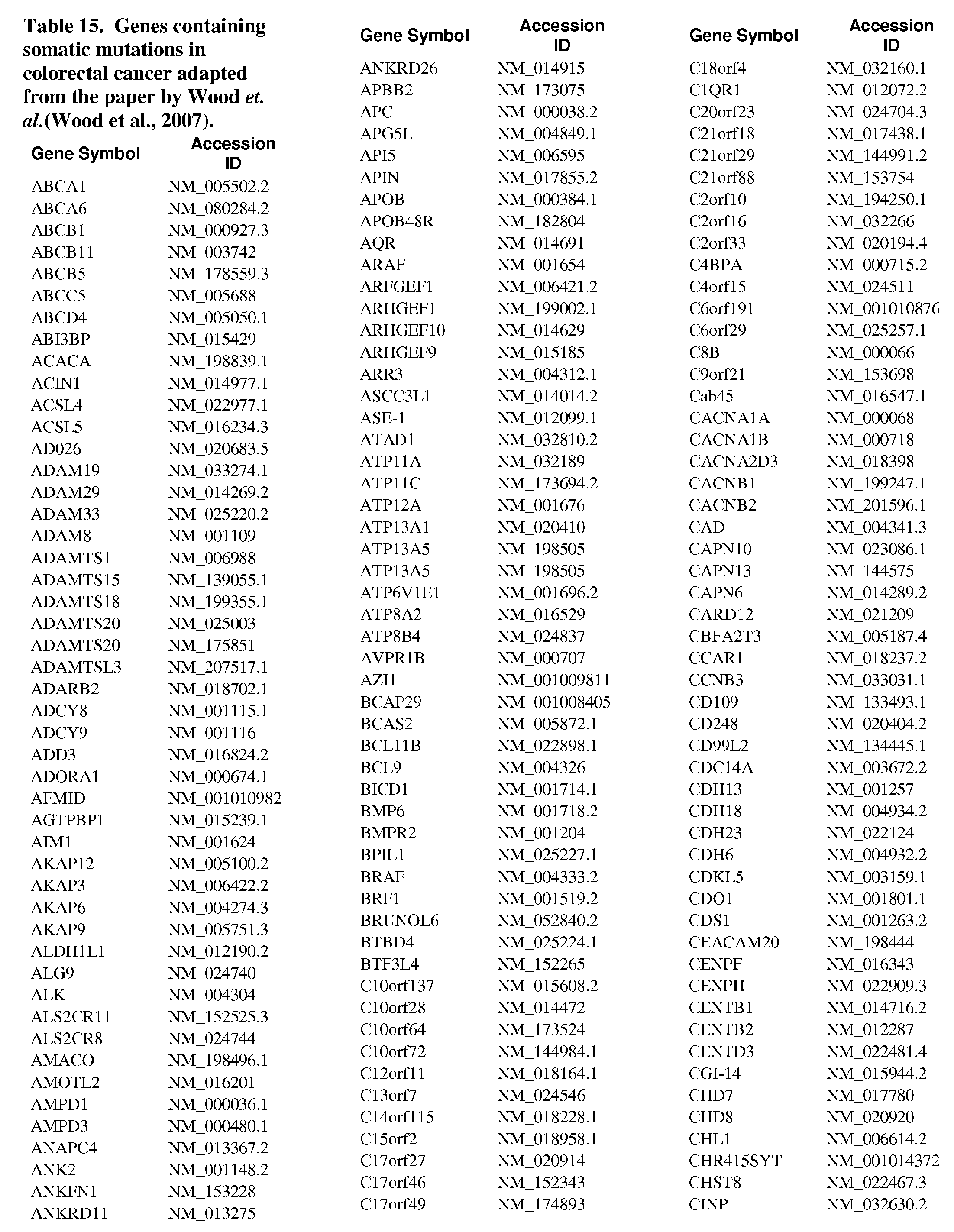


Claims
Priority Applications (28)
| Application Number | Priority Date | Filing Date | Title |
|---|---|---|---|
| EP20161635.6A EP3708682B1 (en) | 2008-02-01 | 2009-02-02 | Use of microvesicles in diagnosis and prognosis of medical diseases and conditions |
| JP2010545256A JP5676277B2 (en) | 2008-02-01 | 2009-02-02 | Use of microvesicles in the diagnosis, prognosis and treatment of medical diseases and conditions |
| EP22208959.1A EP4219762A1 (en) | 2008-02-01 | 2009-02-02 | Use of microvesicles in diagnosis and prognosis of medical diseases and conditions |
| KR1020177023757A KR101970908B1 (en) | 2008-02-01 | 2009-02-02 | Use of microvesicles in diagnosis, prognosis and treatment of medical diseases and conditions |
| AU2009212543A AU2009212543B2 (en) | 2008-02-01 | 2009-02-02 | Use of microvesicles in diagnosis, prognosis and treatment of medical diseases and conditions |
| ES09708907.2T ES2446301T3 (en) | 2008-02-01 | 2009-02-02 | Use of microvesicles in the diagnosis, prognosis and treatment of diseases and medical conditions |
| CA2713909A CA2713909C (en) | 2008-02-01 | 2009-02-02 | Use of microvesicles in diagnosis, prognosis and treatment of medical diseases and conditions |
| BRPI0907050-8A BRPI0907050B1 (en) | 2008-02-01 | 2009-02-02 | Method for detecting in a subject the presence or absence of a predetermined biomarker nucleic acid associated with prostate cancer |
| KR1020167006659A KR101810799B1 (en) | 2008-02-01 | 2009-02-02 | Use of microvesicles in diagnosis, prognosis and treatment of medical diseases and conditions |
| DK09708907.2T DK2245199T3 (en) | 2008-02-01 | 2009-02-02 | APPLICATION OF MICROVESCIES IN DIAGNOSTICATION, PROGRAMMING AND TREATMENT OF MEDICAL DISEASES AND CONDITIONS |
| EP09708907.2A EP2245199B1 (en) | 2008-02-01 | 2009-02-02 | Use of microvesicles in diagnosis, prognosis and treatment of medical diseases and conditions |
| US12/865,681 US20110053157A1 (en) | 2008-02-01 | 2009-02-02 | Use of microvesicles in diagnosis, prognosis and treatment of medical diseases and conditions |
| CN200980111107.0A CN102084000B (en) | 2008-02-01 | 2009-02-02 | The purposes of microvesicle in the diagnosis of medical conditions and the patient's condition, prognosis and treatment |
| EP13151745.0A EP2604704B1 (en) | 2008-02-01 | 2009-02-02 | Use of microvesicles in diagnosis and prognosis of brain tumor |
| US12/695,910 US20100196426A1 (en) | 2008-02-01 | 2010-01-28 | Use of microvesicles in diagnosis and prognosis of medical diseases and conditions |
| US12/727,135 US20110003704A1 (en) | 2008-02-01 | 2010-03-18 | Use of microvesicles in diagnosis and prognosis of medical diseases and conditions |
| US13/294,144 US20120142001A1 (en) | 2008-02-01 | 2011-11-10 | Method for isolation of nucleic acid containing particles and extraction of nucleic acids therefrom |
| HK11113062.3A HK1158705A1 (en) | 2008-02-01 | 2011-12-01 | Use of microvesicles in diagnosis, prognosis and treatment of medical diseases and conditions |
| US13/688,273 US20130131194A1 (en) | 2008-02-01 | 2012-11-29 | Use of microvesicles in diagnosis and prognosis of medical diseases and conditions |
| US13/939,242 US20140194613A1 (en) | 2008-02-01 | 2013-07-11 | Use of microvesicles in diagnosis and prognosis of medical diseases and conditions |
| US14/219,399 US20140194319A1 (en) | 2008-02-01 | 2014-03-19 | Use of microvesicles in diagnosis and prognosis of medical diseases and conditions |
| AU2015203111A AU2015203111B2 (en) | 2008-02-01 | 2015-06-11 | Use of microvesicles in diagnosis, prognosis and treatment of medical diseases and conditions |
| US14/924,245 US20160362678A1 (en) | 2008-02-01 | 2015-10-27 | Use of microvesicles in diagnosis and prognosis of medical diseases and conditions |
| US15/374,477 US20180051335A9 (en) | 2008-02-01 | 2016-12-09 | Use of microvesicles in diagnosis and prognosis of medical diseases and conditions |
| US15/587,953 US20170314075A1 (en) | 2008-02-01 | 2017-05-05 | Use of microvesicles in diagnosis and prognosis of medical diseases and conditions |
| AU2017272272A AU2017272272A1 (en) | 2008-02-01 | 2017-12-07 | Use of microvesicles in diagnosis, prognosis and treatment of medical diseases and conditions |
| AU2020201821A AU2020201821B2 (en) | 2008-02-01 | 2020-03-12 | Use of microvesicles in diagnosis, prognosis and treatment of medical diseases and conditions |
| US17/667,132 US20220162704A1 (en) | 2008-02-01 | 2022-02-08 | Methods of preparing microvesicle micrornas from bodily fluids |
Applications Claiming Priority (4)
| Application Number | Priority Date | Filing Date | Title |
|---|---|---|---|
| US2553608P | 2008-02-01 | 2008-02-01 | |
| US61/025,536 | 2008-02-01 | ||
| US10029308P | 2008-09-26 | 2008-09-26 | |
| US61/100,293 | 2008-09-26 |
Related Parent Applications (1)
| Application Number | Title | Priority Date | Filing Date |
|---|---|---|---|
| US12/838,413 Continuation-In-Part US20110195426A1 (en) | 2008-02-01 | 2010-07-16 | Nucleic acids analysis |
Related Child Applications (5)
| Application Number | Title | Priority Date | Filing Date |
|---|---|---|---|
| US12/865,681 A-371-Of-International US20110053157A1 (en) | 2008-02-01 | 2009-02-02 | Use of microvesicles in diagnosis, prognosis and treatment of medical diseases and conditions |
| US12/695,910 Continuation US20100196426A1 (en) | 2008-02-01 | 2010-01-28 | Use of microvesicles in diagnosis and prognosis of medical diseases and conditions |
| US12/727,135 Continuation US20110003704A1 (en) | 2008-02-01 | 2010-03-18 | Use of microvesicles in diagnosis and prognosis of medical diseases and conditions |
| US13/294,144 Continuation-In-Part US20120142001A1 (en) | 2008-02-01 | 2011-11-10 | Method for isolation of nucleic acid containing particles and extraction of nucleic acids therefrom |
| US13/688,273 Continuation US20130131194A1 (en) | 2008-02-01 | 2012-11-29 | Use of microvesicles in diagnosis and prognosis of medical diseases and conditions |
Publications (1)
| Publication Number | Publication Date |
|---|---|
| WO2009100029A1 true WO2009100029A1 (en) | 2009-08-13 |
Family
ID=40566310
Family Applications (1)
| Application Number | Title | Priority Date | Filing Date |
|---|---|---|---|
| PCT/US2009/032881 WO2009100029A1 (en) | 2008-02-01 | 2009-02-02 | Use of microvesicles in diagnosis, prognosis and treatment of medical diseases and conditions |
Country Status (13)
| Country | Link |
|---|---|
| US (9) | US20110053157A1 (en) |
| EP (6) | EP4219762A1 (en) |
| JP (3) | JP5676277B2 (en) |
| KR (3) | KR101810799B1 (en) |
| CN (2) | CN102084000B (en) |
| AU (1) | AU2009212543B2 (en) |
| BR (1) | BRPI0907050B1 (en) |
| CA (1) | CA2713909C (en) |
| DK (1) | DK2245199T3 (en) |
| ES (3) | ES2703363T3 (en) |
| HK (1) | HK1158705A1 (en) |
| SG (2) | SG10201609507TA (en) |
| WO (1) | WO2009100029A1 (en) |
Cited By (139)
| Publication number | Priority date | Publication date | Assignee | Title |
|---|---|---|---|---|
| GB2463401A (en) * | 2008-11-12 | 2010-03-17 | Caris Mpi Inc | Diagnostic methods using exosomes |
| WO2010072410A3 (en) * | 2008-12-23 | 2010-09-10 | Universiteit Leiden | Methods for immobilizing microvesicles, means and methods for detecting them, and uses thereof |
| US7888035B2 (en) | 2008-10-30 | 2011-02-15 | Caris Mpi, Inc. | Methods for assessing RNA patterns |
| WO2011031877A1 (en) * | 2009-09-09 | 2011-03-17 | The General Hospital Corporation | Use of microvesicles in analyzing nucleic acid profiles |
| WO2011044513A1 (en) * | 2009-10-08 | 2011-04-14 | Wake Forest University Health Sciences | Diagnostic and prognostic markers for cancer |
| US7943318B2 (en) | 2006-01-05 | 2011-05-17 | The Ohio State University Research Foundation | Microrna-based methods and compositions for the diagnosis, prognosis and treatment of lung cancer |
| WO2011063324A2 (en) * | 2009-11-20 | 2011-05-26 | The General Hospital Corporation | Microfluidic systems for isolating microvesicles |
| US7985584B2 (en) | 2006-03-20 | 2011-07-26 | The Ohio State University Research Foundation | MicroRNA fingerprints during human megakaryocytopoiesis |
| WO2011090166A1 (en) * | 2010-01-22 | 2011-07-28 | 株式会社医学生物学研究所 | Esophageal cancer marker |
| WO2011107962A1 (en) | 2010-03-03 | 2011-09-09 | Consiglio Nazionale Delle Ricerche | Increase of myeloid microvesicles in the cerebrospinal fluid as biomarker of microglia/macrophage activation in neurological disorders |
| US8034560B2 (en) | 2007-01-31 | 2011-10-11 | The Ohio State University Research Foundation | MicroRNA-based methods and compositions for the diagnosis, prognosis and treatment of acute myeloid leukemia (AML) |
| US8053186B2 (en) | 2007-06-15 | 2011-11-08 | The Ohio State University Research Foundation | Oncogenic ALL-1 fusion proteins for targeting Drosha-mediated microRNA processing |
| WO2011147086A1 (en) * | 2010-05-26 | 2011-12-01 | 江苏命码生物科技有限公司 | Microvesicles carrying small interfering rnas, preparation methods and uses thereof |
| US8071292B2 (en) | 2006-09-19 | 2011-12-06 | The Ohio State University Research Foundation | Leukemia diagnostic methods |
| US8084199B2 (en) | 2006-07-13 | 2011-12-27 | The Ohio State University Research Foundation | Method of diagnosing poor survival prognosis colon cancer using microRNA-21 |
| US20120027749A1 (en) * | 2008-12-15 | 2012-02-02 | Oxford Biomedica (Uk) Limited | Method |
| WO2012031008A2 (en) | 2010-08-31 | 2012-03-08 | The General Hospital Corporation | Cancer-related biological materials in microvesicles |
| WO2012054975A1 (en) * | 2010-10-28 | 2012-05-03 | Clinical Genomics Pty. Ltd. | Method of microvesicle enrichment |
| WO2012064993A1 (en) | 2010-11-10 | 2012-05-18 | Exosome Diagnosties, Inc. | Method for isolation of nucleic acid containing particles and extraction of nucleic acids therefrom |
| EP2454588A1 (en) * | 2009-07-16 | 2012-05-23 | The General Hospital Corporation | Nucleic acid analysis |
| EP2475989A1 (en) * | 2009-09-09 | 2012-07-18 | The General Hospital Corporation | Use of microvesicles in analyzing kras mutations |
| US8252538B2 (en) | 2006-11-01 | 2012-08-28 | The Ohio State University | MicroRNA expression signature for predicting survival and metastases in hepatocellular carcinoma |
| WO2012135844A2 (en) * | 2011-04-01 | 2012-10-04 | Cornell University | Circulating exosomes as diagnostic/prognostic indicators and therapeutic targets of melanoma and other cancers |
| WO2012155014A1 (en) | 2011-05-11 | 2012-11-15 | Exosome Diagnostics, Inc. | Nucleic acid extraction from heterogeneous biological materials |
| WO2012020307A3 (en) * | 2010-08-13 | 2012-11-29 | The University Court Of The University Of Glasgow | Therapeutic uses of microvesicles and related micrornas |
| US20130012409A1 (en) * | 2009-10-08 | 2013-01-10 | M Frank | Diagnostic and Prognostic Markers for Cancer |
| US8367632B2 (en) | 2007-07-31 | 2013-02-05 | Ohio State University Research Foundation | Methods for reverting methylation by targeting methyltransferases |
| WO2013028788A1 (en) | 2011-08-22 | 2013-02-28 | Exosome Diagnostics, Inc. | Urine biomarkers |
| US8389210B2 (en) | 2006-01-05 | 2013-03-05 | The Ohio State University Research Foundation | MicroRNA expression abnormalities in pancreatic endocrine and acinar tumors |
| US20130065769A1 (en) * | 2011-09-08 | 2013-03-14 | The Regents Of The University Of California | Salivary biomarkers for gastric cancer detection |
| JP2013524247A (en) * | 2010-04-13 | 2013-06-17 | ザ ロイヤル インスティチューション フォー ザ アドバンスメント オブ ラーニング/マクギル ユニバーシティ | Tumor cell-derived microvesicles |
| US8465917B2 (en) | 2007-06-08 | 2013-06-18 | The Ohio State University Research Foundation | Methods for determining heptocellular carcinoma subtype and detecting hepatic cancer stem cells |
| US8466119B2 (en) | 2007-08-22 | 2013-06-18 | The Ohio State University Research Foundation | Methods and compositions for inducing deregulation of EPHA7 and ERK phosphorylation in human acute leukemias |
| EP2606353A1 (en) * | 2010-08-18 | 2013-06-26 | Caris Life Sciences Luxembourg Holdings | Circulating biomarkers for disease |
| JP2013528063A (en) * | 2010-06-11 | 2013-07-08 | 日立化成株式会社 | How to determine the characteristics of renal function |
| JP2013528375A (en) * | 2010-05-12 | 2013-07-11 | テンジオン, インク. | Bioactive kidney cells |
| EP2627788A2 (en) * | 2010-10-15 | 2013-08-21 | The General Hospital Corporation | Microvesicle-based assays |
| JP2013534830A (en) * | 2010-07-01 | 2013-09-09 | イオン メディックス インコーポレイテッド | Microvesicles derived from cell protoplasts and uses thereof |
| CN103374625A (en) * | 2012-04-27 | 2013-10-30 | 中国科学院上海生命科学研究院 | Congenital heart disease related gene DLC1 and application thereof |
| CN103477225A (en) * | 2011-02-17 | 2013-12-25 | 康奈尔大学 | Compositions and methods to prevent cell transformation and cancer metastasis |
| US8658370B2 (en) | 2005-08-01 | 2014-02-25 | The Ohio State University Research Foundation | MicroRNA-based methods and compositions for the diagnosis, prognosis and treatment of breast cancer |
| EP2699666A1 (en) * | 2011-04-18 | 2014-02-26 | DiamiR, LLC | miRNA-BASED UNIVERSAL SCREENING TEST (UST) |
| US8664192B2 (en) | 2011-03-07 | 2014-03-04 | The Ohio State University | Mutator activity induced by microRNA-155 (miR-155) links inflammation and cancer |
| WO2014041087A1 (en) | 2012-09-12 | 2014-03-20 | INSERM (Institut National de la Santé et de la Recherche Médicale) | Muscle secretome and uses thereof |
| JP2014507140A (en) * | 2011-02-11 | 2014-03-27 | エイジェンシー・フォー・サイエンス,テクノロジー・アンド・リサーチ | Methods for detecting therapeutic exosomes |
| WO2014057128A2 (en) | 2012-10-11 | 2014-04-17 | Vin-De-Bona Trading Company Pte Ltd | Method of painting microvesicles |
| WO2014072468A1 (en) * | 2012-11-09 | 2014-05-15 | Velin-Pharma A/S | Compositions for pulmonary delivery |
| US20140275211A1 (en) * | 2011-06-21 | 2014-09-18 | Alnylam Pharmaceuticals, Inc. | Assays and methods for determining activity of a therapeutic agent in a subject |
| US8859202B2 (en) | 2012-01-20 | 2014-10-14 | The Ohio State University | Breast cancer biomarker signatures for invasiveness and prognosis |
| US8911998B2 (en) | 2007-10-26 | 2014-12-16 | The Ohio State University | Methods for identifying fragile histidine triad (FHIT) interaction and uses thereof |
| US8916533B2 (en) | 2009-11-23 | 2014-12-23 | The Ohio State University | Materials and methods useful for affecting tumor cell growth, migration and invasion |
| WO2015005553A1 (en) * | 2013-07-12 | 2015-01-15 | Kwon Young Ah | Method for diagnosis of diseases using morphological characteristics of luterial |
| US8946187B2 (en) | 2010-11-12 | 2015-02-03 | The Ohio State University | Materials and methods related to microRNA-21, mismatch repair, and colorectal cancer |
| CN104372004A (en) * | 2014-12-04 | 2015-02-25 | 广东医学院 | Detection method and application of single-nucleotide polymorphic sites of miR-27a gene associated with myocardial infarction susceptibility |
| CN104404044A (en) * | 2014-12-04 | 2015-03-11 | 广东医学院 | Detection method and applications for single nucleotide polymorphisms in exon region of ANRIL (Antisense Noncoding RNA in the INK4 Locus) gene related to susceptibility of coronary artery disease |
| EP2670868A4 (en) * | 2011-01-31 | 2015-05-06 | Esoterix Genetic Lab Llc | Methods for enriching microparticles or nucleic acids using binding molecules |
| US9085804B2 (en) | 2007-08-03 | 2015-07-21 | The Ohio State University Research Foundation | Ultraconserved regions encoding ncRNAs |
| US9128101B2 (en) | 2010-03-01 | 2015-09-08 | Caris Life Sciences Switzerland Holdings Gmbh | Biomarkers for theranostics |
| US9125923B2 (en) | 2008-06-11 | 2015-09-08 | The Ohio State University | Use of MiR-26 family as a predictive marker for hepatocellular carcinoma and responsiveness to therapy |
| US9186405B2 (en) | 2007-08-16 | 2015-11-17 | The Royal Institution For The Advancement Of Learning/Mcgill University | Tumor cell-derived microvesicles |
| US9249468B2 (en) | 2011-10-14 | 2016-02-02 | The Ohio State University | Methods and materials related to ovarian cancer |
| EP2875158A4 (en) * | 2012-07-18 | 2016-03-23 | Exosome Diagnostics Inc | Use of microvesicles in diagnosis, prognosis, and treatment of medical diseases and conditions |
| US9421167B2 (en) | 2010-05-26 | 2016-08-23 | Micromedmark Biotech Co. Ltd. | Preparation of microvesicle-siRNA complexes and use thereof in AIDS treatment |
| US9458496B2 (en) | 2010-06-11 | 2016-10-04 | Hitachi Chemical Company, Ltd. | Method of characterizing vascular diseases |
| US9469876B2 (en) | 2010-04-06 | 2016-10-18 | Caris Life Sciences Switzerland Holdings Gmbh | Circulating biomarkers for metastatic prostate cancer |
| US9481885B2 (en) | 2011-12-13 | 2016-11-01 | Ohio State Innovation Foundation | Methods and compositions related to miR-21 and miR-29a, exosome inhibition, and cancer metastasis |
| EP2875348A4 (en) * | 2012-07-19 | 2016-11-02 | Atlantic Cancer Res Inst | Method for the isolation of microvesicles |
| EP2961851A4 (en) * | 2013-02-28 | 2016-11-16 | Univ Hong Kong Chinese | Maternal plasma transcriptome analysis by massively parallel rna sequencing |
| US9662649B2 (en) | 2013-05-06 | 2017-05-30 | Hitachi Chemical Company America, Ltd. | Devices and methods for capturing target molecules |
| US9719129B2 (en) | 2011-06-10 | 2017-08-01 | Hitachi Chemical Co., Ltd. | Methods for isolating vesicles from biological fluids |
| WO2017181183A1 (en) * | 2016-04-15 | 2017-10-19 | Exosome Diagnostics, Inc. | Plasma-based detection of anaplastic lymphoma kinase (alk) nucleic acids and alk fusion transcripts and uses thereof in diagnosis and treatment of cancer |
| WO2017197399A1 (en) | 2016-05-13 | 2017-11-16 | Exosome Diagnostics, Inc. | Automated and manual methods for isolation of extracellular vesicles and co-isolation of cell-free dna from biofluids |
| EP3125907A4 (en) * | 2014-04-01 | 2017-11-29 | Cornell University | Use of double-stranded dna in exosomes: a novel biomarker in cancer detection |
| EP3095875A4 (en) * | 2014-01-14 | 2018-01-03 | Won Cheol Choi | Luterial and method for isolating and culturing same |
| AU2011320021B2 (en) * | 2011-10-26 | 2018-02-22 | Clinical Genomics Pty. Ltd. | Method of microvesicle enrichment |
| WO2018076018A1 (en) | 2016-10-21 | 2018-04-26 | Exosome Diagnostics, Inc. | Sequencing and analysis of exosome associated nucleic acids |
| EP3243902A4 (en) * | 2015-01-06 | 2018-05-30 | Luterion Co., Ltd. | Luterion and separating and culturing methods for same |
| WO2018106648A1 (en) * | 2016-12-05 | 2018-06-14 | The Penn State Research Foundation | Lipid-based probes for extracellular isolation |
| WO2018126278A2 (en) | 2017-01-02 | 2018-07-05 | Exosome Diagnostics, Inc. | Methods to distinguish rna and dna in a combined preparation |
| WO2018143896A1 (en) * | 2017-02-02 | 2018-08-09 | Agency For Science, Technology And Research | Ovarian cancer biomarker |
| US10071170B2 (en) | 2013-06-24 | 2018-09-11 | Ablbio | Antibody-drug conjugate having improved stability and use thereof |
| WO2018195308A1 (en) * | 2017-04-19 | 2018-10-25 | Figene, Llc | Stimulation of angiogenesis by fibroblast derived exosomes |
| US10174361B2 (en) | 2011-11-10 | 2019-01-08 | Exosome Diagnostics, Inc. | Cerebrospinal fluid assay |
| US10260067B2 (en) * | 2014-10-01 | 2019-04-16 | The Brigham And Women's Hospital, Inc. | Enhancing dermal wound healing by downregulating microRNA-26a |
| US10266895B2 (en) | 2014-11-05 | 2019-04-23 | Hitachi Chemical Company Ltd. | Exosomes and microvesicles in intestinal luminal fluids and stool and use of same for the assessment of inflammatory bowel disease |
| AU2013340414B2 (en) * | 2012-10-23 | 2019-05-02 | Caris Science, Inc. | Aptamers and uses thereof |
| US10301681B2 (en) | 2013-08-06 | 2019-05-28 | Exosome Diagnostics, Inc. | Methods of treating a subject with a high gleason score prostate cancer |
| EP3499212A1 (en) | 2013-01-03 | 2019-06-19 | Exosome Diagnostics Inc. | Methods for isolating microvesicles |
| US10370719B2 (en) | 2014-11-12 | 2019-08-06 | Hitachi Chemical Co., Ltd. | Method and device for diagnosing organ injury |
| US10378060B2 (en) | 2011-10-14 | 2019-08-13 | Dana-Farber Cancer Institute, Inc. | ZNF365/ZFP365 biomarker predictive of anti-cancer response |
| US10465183B2 (en) | 2014-07-09 | 2019-11-05 | Exosome Diagnostics, Inc. | Methods for isolating microvesicles and extracting nucleic acids from biological samples |
| CN110546278A (en) * | 2017-02-24 | 2019-12-06 | Md保健株式会社 | Method for diagnosing chronic obstructive respiratory disease by bacterial metagenomic analysis |
| WO2019236853A1 (en) | 2018-06-06 | 2019-12-12 | Exosome Diagnostics, Inc. | Methods for developing urine biomarkers and for detecting bladder cancer |
| EP3427060A4 (en) * | 2016-03-07 | 2019-12-18 | CFGenome, LLC | Noninvasive molecular controls |
| US10590384B2 (en) | 2014-01-14 | 2020-03-17 | Luterion Co., Ltd. | Luterial and method for isolating and culturing the same |
| US10598665B2 (en) | 2013-12-04 | 2020-03-24 | Board Of Regents, The University Of Texas System | Analysis of genomic DNA, RNA, and proteins in exosomes for diagnosis and theranosis |
| US10607717B2 (en) | 2013-11-06 | 2020-03-31 | The United States Of America, As Represented By The Secretary, Department Of Health And Human Services | Method for subtyping lymphoma types by means of expression profiling |
| WO2020106853A1 (en) | 2018-11-20 | 2020-05-28 | Exosome Diagnostics, Inc. | Compositions and methods for internal controls of microvesicle isolations |
| US10758619B2 (en) | 2010-11-15 | 2020-09-01 | The Ohio State University | Controlled release mucoadhesive systems |
| EP3564388A4 (en) * | 2016-12-28 | 2020-09-02 | MD Healthcare Inc. | Method for diagnosing prostatic disease via bacterial metagenomic analysis |
| US10781487B2 (en) | 2017-07-24 | 2020-09-22 | Diamir, Llc | miRNA-based methods for detecting and monitoring aging |
| US10914748B2 (en) | 2016-09-08 | 2021-02-09 | UNIVERSITé LAVAL | Erythrocyte-derived extracellular vesicles as a biomarker for clinically assessing Parkinson's disease |
| US10975436B2 (en) | 2016-01-05 | 2021-04-13 | Diamir, Llc | Methods of using miRNA from bodily fluids for diagnosis and monitoring of neurodevelopmental disorders |
| US11028443B2 (en) | 2015-08-31 | 2021-06-08 | Showa Denko Materials Co., Ltd. | Molecular methods for assessing urothelial disease |
| US11098362B2 (en) | 2013-11-18 | 2021-08-24 | Diamir, Llc | Methods of using miRNAs from bodily fluids for detection and monitoring of Parkinson's disease (PD) |
| EP3655531A4 (en) * | 2017-07-18 | 2021-09-08 | Exosome Diagnostics, Inc. | Sequencing of nucleic acids associated with exosomal isolation from patients with glioblastoma multiforme |
| US11136627B2 (en) | 2012-08-30 | 2021-10-05 | Exosome Diagnostics, Inc. | Controls for nucleic acid assays |
| US11149313B2 (en) | 2016-03-21 | 2021-10-19 | Diamir, Llc | Methods of using miRNAs from bodily fluids for detection and differentiation of neurodegenerative diseases |
| US11174503B2 (en) | 2016-09-21 | 2021-11-16 | Predicine, Inc. | Systems and methods for combined detection of genetic alterations |
| US11180812B2 (en) | 2012-08-17 | 2021-11-23 | Cornell University | Use of DNA in circulating exosomes as a diagnostic marker for metastatic disease |
| WO2021243206A1 (en) | 2020-05-29 | 2021-12-02 | Exosome Diagnostics, Inc. | Use of microvesicle signature for the diagnosis and treatment of kidney transplant rejection |
| US11266736B2 (en) | 2008-04-17 | 2022-03-08 | Vin De Bona Trading Company Pte Ltd | Method of painting micro vesicles |
| US11268085B2 (en) | 2014-05-27 | 2022-03-08 | Exosome Diagnostics, Inc. | Methods for isolating microvesicles and extracting nucleic acids from biological samples |
| CN114231643A (en) * | 2022-01-07 | 2022-03-25 | 新疆畜牧科学院畜牧研究所 | Molecular marker influencing character of diameter variation coefficient of Eldos fine wool sheep wool, obtaining method, specific primer pair and application thereof |
| US11286530B2 (en) | 2010-05-18 | 2022-03-29 | Natera, Inc. | Methods for simultaneous amplification of target loci |
| US11306359B2 (en) | 2005-11-26 | 2022-04-19 | Natera, Inc. | System and method for cleaning noisy genetic data from target individuals using genetic data from genetically related individuals |
| US11306357B2 (en) | 2010-05-18 | 2022-04-19 | Natera, Inc. | Methods for non-invasive prenatal ploidy calling |
| US11319595B2 (en) | 2014-04-21 | 2022-05-03 | Natera, Inc. | Detecting mutations and ploidy in chromosomal segments |
| US11322224B2 (en) | 2010-05-18 | 2022-05-03 | Natera, Inc. | Methods for non-invasive prenatal ploidy calling |
| US11326208B2 (en) | 2010-05-18 | 2022-05-10 | Natera, Inc. | Methods for nested PCR amplification of cell-free DNA |
| US11332785B2 (en) | 2010-05-18 | 2022-05-17 | Natera, Inc. | Methods for non-invasive prenatal ploidy calling |
| US11332793B2 (en) | 2010-05-18 | 2022-05-17 | Natera, Inc. | Methods for simultaneous amplification of target loci |
| US11339429B2 (en) | 2010-05-18 | 2022-05-24 | Natera, Inc. | Methods for non-invasive prenatal ploidy calling |
| US11390916B2 (en) | 2014-04-21 | 2022-07-19 | Natera, Inc. | Methods for simultaneous amplification of target loci |
| US11397182B2 (en) | 2014-10-07 | 2022-07-26 | Cornell University | Methods for prognosing and preventing metastatic liver disease |
| US11408031B2 (en) | 2010-05-18 | 2022-08-09 | Natera, Inc. | Methods for non-invasive prenatal paternity testing |
| US11479812B2 (en) | 2015-05-11 | 2022-10-25 | Natera, Inc. | Methods and compositions for determining ploidy |
| US11485996B2 (en) | 2016-10-04 | 2022-11-01 | Natera, Inc. | Methods for characterizing copy number variation using proximity-litigation sequencing |
| US11519028B2 (en) | 2016-12-07 | 2022-12-06 | Natera, Inc. | Compositions and methods for identifying nucleic acid molecules |
| US11519035B2 (en) | 2010-05-18 | 2022-12-06 | Natera, Inc. | Methods for simultaneous amplification of target loci |
| US11525159B2 (en) | 2018-07-03 | 2022-12-13 | Natera, Inc. | Methods for detection of donor-derived cell-free DNA |
| US11702702B2 (en) | 2016-04-15 | 2023-07-18 | Predicine, Inc. | Systems and methods for detecting genetic alterations |
| WO2023158869A1 (en) | 2022-02-18 | 2023-08-24 | Exosome Diagnostics, Inc. | Use of microvesicle signatures in the identification and treatment of renal disorders |
| US11752175B2 (en) | 2018-09-21 | 2023-09-12 | Cornell University | Compositions and methods for glaucoma |
| EP4006166A4 (en) * | 2019-09-05 | 2023-09-27 | Nanjing Anji Biotechnology Co., Ltd. | Tumour marker aquaporin 2 protein and application thereof |
| US11891664B2 (en) | 2017-05-17 | 2024-02-06 | Exosome Diagnostics, Inc. | Methods of detecting renal transplant rejection using microvesicle nucleic acid biomarkers |
| US11899024B2 (en) | 2017-07-12 | 2024-02-13 | Exosome Diagnostics, Inc. | Treatment and diagnosis of parkinson's disease using isolated and enriched populations of biofluid-derived extracellular vesicles |
| WO2024054572A1 (en) | 2022-09-07 | 2024-03-14 | Exosome Diagnostics, Inc. | Methods of detecting sjögren's syndrome using salivary exosomes |
| US11939634B2 (en) | 2010-05-18 | 2024-03-26 | Natera, Inc. | Methods for simultaneous amplification of target loci |
Families Citing this family (165)
| Publication number | Priority date | Publication date | Assignee | Title |
|---|---|---|---|---|
| US8021847B2 (en) | 2004-06-02 | 2011-09-20 | Proxy Life Science Holdings, Inc. | Microvesicle-based compositions and methods |
| WO2005121369A2 (en) * | 2004-06-02 | 2005-12-22 | Sourcepharm, Inc. | Rna-containing microvesicles and methods therefor |
| JP2008538700A (en) | 2005-04-22 | 2008-11-06 | モルフォテック、インク. | Antibodies internalized into endosialin cells with immune effector activity |
| AU2006291165B2 (en) | 2005-09-12 | 2013-03-14 | The Ohio State University Research Foundation | Compositions and methods for the diagnosis and therapy of BCL2-associated cancers |
| EP2487252B1 (en) | 2006-01-05 | 2014-10-15 | The Ohio State University Research Foundation | MicroRNA-based methods for the diagnosis of colon cancer |
| ES2880460T3 (en) * | 2006-03-09 | 2021-11-24 | Aethlon Medical Inc | Extracorporeal removal of microvesicular particles |
| LT2620451T (en) * | 2007-04-05 | 2017-01-10 | Morphotek, Inc. | Methods for inhibiting the binding of endosialin to ligands |
| JP5592251B2 (en) * | 2007-04-30 | 2014-09-17 | ジ・オハイオ・ステイト・ユニバーシティ・リサーチ・ファウンデイション | Method for distinguishing pancreatic cancer from normal pancreatic function and / or chronic pancreatitis |
| AU2008310704B2 (en) * | 2007-10-11 | 2014-03-20 | The Government Of The United States Of America As Represented By The Secretary Of The Department Of Health And Human Services | Methods and compositions for the diagnosis and treatment of esphageal adenocarcinomas |
| US20120058492A1 (en) * | 2008-01-25 | 2012-03-08 | Hansabiomed Ou | Method and a Kit To Detect Malignant Tumors and Provide a Prognosis |
| US20110054009A1 (en) * | 2008-02-28 | 2011-03-03 | The Ohio State University Research Foundation | MicroRNA-Based Methods and Compositions for the Diagnosis, Prognosis and Treatment of Prostate Related Disorders |
| JP5662166B2 (en) * | 2008-02-28 | 2015-01-28 | ジ・オハイオ・ステイト・ユニバーシティ・リサーチ・ファウンデイションThe Ohio State University Research Foundation | MicroRNA-based methods and compositions for diagnosis, prognosis and treatment of gastric cancer |
| JP2012500389A (en) * | 2008-08-12 | 2012-01-05 | ジ・オハイオ・ステイト・ユニバーシティ・リサーチ・ファウンデイション | MicroRNA-based compositions and methods for diagnosis, prognosis and treatment of multiple myeloma |
| AU2009288097B2 (en) * | 2008-09-03 | 2013-11-14 | The Johns Hopkins University | Pathways underlying pancreatic tumorigenesis and an hereditary pancreatic cancer gene |
| US9487837B2 (en) | 2008-10-06 | 2016-11-08 | Morehouse School Of Medicine | Exosome-mediated diagnosis of hepatitis virus infections and diseases |
| US20110301221A1 (en) * | 2008-10-10 | 2011-12-08 | Swedish Health Services | Diagnosis, prognosis and treatment of glioblastoma multiforme |
| WO2011006119A2 (en) | 2009-07-09 | 2011-01-13 | The Scripps Research Institute | Gene expression profiles associated with chronic allograft nephropathy |
| AU2010324594B2 (en) * | 2009-11-30 | 2016-09-15 | Caris Life Sciences Switzerland Holdings Gmbh | Methods and systems for isolating, storing, and analyzing vesicles |
| US9315802B2 (en) | 2009-12-30 | 2016-04-19 | Quest Diagnostics Investments Incorporated | RNA isolation from soluble urine fractions |
| EP2848703A1 (en) | 2010-01-19 | 2015-03-18 | Verinata Health, Inc | Simultaneous determination of aneuploidy and fetal fraction |
| US20120100548A1 (en) | 2010-10-26 | 2012-04-26 | Verinata Health, Inc. | Method for determining copy number variations |
| US20130052647A1 (en) * | 2010-02-10 | 2013-02-28 | Marianna Goldrick | Methods for fractionating and processing microparticles from biological samples and using them for biomarker discovery |
| EP2415877A3 (en) * | 2010-02-17 | 2012-02-15 | Deutsches Krebsforschungszentrum | Means and methods for diagnosing pancreatic cancer |
| EP2558597A4 (en) * | 2010-04-15 | 2013-11-20 | Hadasit Med Res Service | Early detection and staging of colorectal cancer using a panel of micro rnas |
| WO2011140662A1 (en) * | 2010-05-13 | 2011-11-17 | The Royal Institution For The Advancement Of Learning / Mcgill University | Cux1 signature for determination of cancer clinical outcome |
| WO2012017430A2 (en) | 2010-08-01 | 2012-02-09 | Tel Hashomer Medical Research Infrastructure And Services Ltd. | Microrna patterns for the diagnosis, prognosis and treatment of melanoma |
| US20120053062A1 (en) * | 2010-08-24 | 2012-03-01 | Bio Dx, Inc. | Defining diagnostic and therapeutic targets of conserved free floating fetal dna in maternal circulating blood |
| WO2012071096A2 (en) * | 2010-09-03 | 2012-05-31 | The Johns Hopkins University | Arid1a and ppp2r1a mutations in cancer |
| EP2619329B1 (en) | 2010-09-24 | 2019-05-22 | The Board of Trustees of The Leland Stanford Junior University | Direct capture, amplification and sequencing of target dna using immobilized primers |
| CA3065694A1 (en) | 2010-11-10 | 2012-05-18 | Inregen | Methods of forming injectable formulations for providing regenerative effects to an organ such as a kidney |
| WO2012067899A2 (en) * | 2010-11-15 | 2012-05-24 | Vanderbilt University | Foxa1 as a marker for invasive bladder cancer |
| US9308255B2 (en) | 2010-11-24 | 2016-04-12 | Yale University | Methods of administering D-dopachrome tautomerase (D-DT) to treat ischemia-reperfusion injury |
| WO2012092490A1 (en) | 2010-12-30 | 2012-07-05 | Quest Diagnostics Investments Incorporated | Diagnosis of prostate cancer |
| WO2012097047A1 (en) * | 2011-01-11 | 2012-07-19 | The Ohio State University Research Foundation | Methods to identify chronic lymphocytic leukemia disease progression |
| WO2012100023A1 (en) * | 2011-01-19 | 2012-07-26 | Thomas Jefferson University | Detection of pancreatic ductal adenocarcinoma |
| WO2012106338A1 (en) * | 2011-01-31 | 2012-08-09 | Esoterix Genetic Laboratories, Llc | Methods for enriching microparticles or nucleic acids in a complex mixture using size exclusion filtration |
| EP2484779A1 (en) | 2011-02-03 | 2012-08-08 | Medizinische Hochschule Hannover | Device and method for analysis of kidney failure |
| WO2012135814A2 (en) * | 2011-03-31 | 2012-10-04 | University Of Houston | Microrna29a,b,c as a tumor suppressor and sensitizing agent for chemotherapy |
| EP2718721A4 (en) * | 2011-06-07 | 2014-10-01 | Caris Life Sciences Luxembourg Holdings S A R L | Circulating biomarkers for cancer |
| EP2739754A4 (en) * | 2011-08-05 | 2015-01-21 | Univ Louisville Res Found | Microrna biomarkers |
| WO2013023376A1 (en) * | 2011-08-18 | 2013-02-21 | Ming-Chung Jiang | Microvesicle membrane protein and application thereof |
| WO2013041913A1 (en) * | 2011-09-22 | 2013-03-28 | Universidad De Los Andes | Method for monitoring, diagnosis and/or prognosis of acute kidney injury in early stage |
| WO2013048734A1 (en) * | 2011-09-28 | 2013-04-04 | Tufts Medical Center, Inc. | Treatment and prevention of cardiovascular disease with cell derived lipid vesicles, microvesicles and exosomes |
| FR2981662A1 (en) * | 2011-10-20 | 2013-04-26 | Assist Publ Hopitaux De Paris | METHOD OF QUANTIFYING RENAL MARKERS BY URINARY DOSAGE. |
| WO2013063439A1 (en) * | 2011-10-27 | 2013-05-02 | University Of Louisville Research Foundation, Inc. | Characterizing melanoma |
| WO2013067048A1 (en) * | 2011-10-31 | 2013-05-10 | The Ohio State University | Materials and methods related to mir-221 and hepatocellular carcinoma |
| ITRM20110685A1 (en) | 2011-12-23 | 2013-06-24 | Internat Ct For Genetic En Gineering And | MICRORNA FOR CARDIAC REGENERATION THROUGH THE INDUCTION OF THE PROLIFERATION OF CARDIAC MYCYCLES |
| US9737480B2 (en) | 2012-02-06 | 2017-08-22 | President And Fellows Of Harvard College | ARRDC1-mediated microvesicles (ARMMs) and uses thereof |
| US20150152474A1 (en) * | 2012-03-09 | 2015-06-04 | Caris Life Sciences Switzerland Holdings Gmbh | Biomarker compositions and methods |
| WO2013148147A1 (en) | 2012-03-26 | 2013-10-03 | The U.S.A., As Represented By The Secretary Dept. Of Health And Human Services | Dna methylation analysis for the diagnosis, prognosis and treatment of adrenal neoplasms |
| US9334498B2 (en) | 2012-05-10 | 2016-05-10 | Uab Research Foundation | Methods and compositions for modulating MIR-204 activity |
| ES2638944T3 (en) * | 2012-05-21 | 2017-10-24 | The Penn State Research Foundation | Compositions and methods related to variant 2 of dennd1a and polycystic ovary syndrome |
| WO2013184905A1 (en) | 2012-06-06 | 2013-12-12 | Myriad Genetics, Inc. | Hereditary cancer genes |
| US9989539B2 (en) | 2012-06-26 | 2018-06-05 | Temple University—Of the Commonwealth System of Higher Education | Method for detecting injury to the brain |
| NZ703411A (en) | 2012-06-27 | 2017-09-29 | Berg Llc | Use of markers in the diagnosis and treatment of prostate cancer |
| US20140018299A1 (en) * | 2012-07-10 | 2014-01-16 | Banyan Biomarkers, Inc. | Method and device to detect, monitor and promote neural regeneration and improvement of cognitive function in a subject suffering from neural injury |
| US20150252428A1 (en) * | 2012-10-03 | 2015-09-10 | Exosome Diagnostics, Inc. | Use of microvesicles in diagnosis, prognosis, and treatment of medical diseases and conditions |
| TWI485159B (en) * | 2012-10-11 | 2015-05-21 | Ming Chung Jiang | A microvesicle membrane protein and application thereof |
| WO2014062218A1 (en) * | 2012-10-16 | 2014-04-24 | University Of Southern California | Colorectal cancer dna methylation markers |
| US10942184B2 (en) | 2012-10-23 | 2021-03-09 | Caris Science, Inc. | Aptamers and uses thereof |
| US20150246073A1 (en) | 2012-10-24 | 2015-09-03 | Regenmedtx, Llc | Renal cell populations and uses thereof |
| WO2014085666A1 (en) * | 2012-11-27 | 2014-06-05 | Board Of Regents, The University Of Texas System | Methods of characterizing and treating molecular subset of muscle-invasive bladder cancer |
| EP2746406B1 (en) | 2012-12-18 | 2017-08-23 | Samsung Electronics Co., Ltd. | Composition and kit for diagnosing breast cancer including miRNAs within vesicle, and method of diagnosing breast cancer using the same |
| WO2014100434A1 (en) * | 2012-12-19 | 2014-06-26 | Caris Science, Inc. | Compositions and methods for aptamer screening |
| EP2959016A4 (en) * | 2013-02-21 | 2016-10-12 | Toma Biosciences Inc | Methods, compositions, and kits for nucleic acid analysis |
| US10793913B2 (en) * | 2013-03-08 | 2020-10-06 | City Of Hope | Methods and compositions for treatment of breast cancer |
| AU2014244618A1 (en) * | 2013-03-13 | 2015-10-08 | University Of Miami | Method for isolation and purification of microvesicles from cell culture supernatants and biological fluids |
| WO2014160032A1 (en) * | 2013-03-13 | 2014-10-02 | University Of Louisville Research Foundation, Inc. | Exosome-associated rna as a diagnostic marker |
| EP3527670B1 (en) | 2013-03-15 | 2023-10-18 | Beth Israel Deaconess Medical Center, Inc. | Mirna biogenesis in exosomes for diagnosis and therapy |
| KR101473557B1 (en) | 2013-03-29 | 2014-12-24 | 포항공과대학교 산학협력단 | Apparatus for the preparation of cell-derived artificial microvesicles using centrifugal force |
| WO2014166303A2 (en) * | 2013-04-12 | 2014-10-16 | The Chinese University Of Hong Kong | Use of multiomic signature to predict diabetes |
| WO2014172390A2 (en) * | 2013-04-15 | 2014-10-23 | Cedars-Sinai Medical Center | Methods for detecting cancer metastasis |
| US20160097771A1 (en) * | 2013-05-21 | 2016-04-07 | NX Pharmagen | Use of tenascin-c as an extracellular marker of tumor-derived microparticles |
| CN103272248B (en) * | 2013-06-28 | 2016-03-30 | 上海市内分泌代谢病研究所 | The application of miR-15b in preparation treatment of diabetes preparation |
| KR20150043937A (en) | 2013-10-15 | 2015-04-23 | 삼성전자주식회사 | A compositon for diagnosing a liver cancer in a subject, method for diagnosing a liver cancer in a subject and method for obtaining information for diagnosing a liver cancer in a subject |
| TWI708058B (en) * | 2013-10-24 | 2020-10-21 | 美商納諾索米克斯公司 | Biomarkers and diagnostic methods for alzheimer's disease and other neurodegenerative disorders |
| US9676844B2 (en) | 2013-11-19 | 2017-06-13 | Virginia Commonwealth University | Pharmaceutical compositions comprising DENND1A variant 2 and methods of use thereof |
| JP6128621B2 (en) * | 2013-12-04 | 2017-05-17 | 国立研究開発法人産業技術総合研究所 | Nucleic acid aptamers for microvesicles |
| JP2017505448A (en) * | 2014-01-14 | 2017-02-16 | チョイ ウォンチョルCHOI, Won Cheol | Screening method for cancer preventive or anticancer agent using morphological characteristics of ruthereal |
| US20160346334A1 (en) * | 2014-02-05 | 2016-12-01 | Stc.Unm | Exosomes as a therapeutic for cancer |
| ES2824108T3 (en) | 2014-03-12 | 2021-05-11 | Icahn School Med Mount Sinai | Method to identify kidney allograft recipients at risk for chronic injury |
| US10443100B2 (en) | 2014-05-22 | 2019-10-15 | The Scripps Research Institute | Gene expression profiles associated with sub-clinical kidney transplant rejection |
| US11104951B2 (en) | 2014-05-22 | 2021-08-31 | The Scripps Research Institute | Molecular signatures for distinguishing liver transplant rejections or injuries |
| RU2017101174A (en) | 2014-06-18 | 2018-07-18 | Торэй Индастриз, Инк. | KIT OR DEVICE FOR LIVER CANCER DETECTION AND DETECTION METHOD |
| CN106661635B (en) | 2014-06-26 | 2021-05-28 | 西奈山伊坎医学院 | Method for diagnosing subclinical and clinical acute rejection by analyzing predictive gene set |
| US20170227541A1 (en) * | 2014-07-17 | 2017-08-10 | The Trustees Of The University Of Pennsylvania | Methods for using exosomes to monitor transplanted organ status |
| AU2015301150A1 (en) * | 2014-08-05 | 2017-02-23 | Ymir Genomics Llc | Methods for the isolation of extracellular vesicles and other bioparticles from urine and other biofluids |
| US20170298352A1 (en) * | 2014-09-30 | 2017-10-19 | Research Institute at Nationwide Children's Hospit al | Compositions and methods for treating hepatic fibrosis |
| CN104535766B (en) * | 2014-10-30 | 2016-04-13 | 中国人民解放军总医院第一附属医院 | The diagnosing cancer of liver that peripheral blood exosome originates and prognostic marker and application thereof |
| US9816080B2 (en) | 2014-10-31 | 2017-11-14 | President And Fellows Of Harvard College | Delivery of CAS9 via ARRDC1-mediated microvesicles (ARMMs) |
| US20170319649A1 (en) * | 2014-11-19 | 2017-11-09 | The Board of Regents of the University of Texsas System | Novel peptide activator of cyclin c-dependent kinase 8 (cdk8) |
| JP6759229B2 (en) | 2014-12-08 | 2020-09-23 | バーグ エルエルシー | Use of markers containing filamin A in the diagnosis and treatment of prostate cancer |
| EP3034623A1 (en) * | 2014-12-18 | 2016-06-22 | Centro de Investigación Biomédica en Red (CIBER) | Method for predicting response to continuous positive air pressure treatment |
| JP6498274B2 (en) * | 2015-03-12 | 2019-04-10 | 三井化学株式会社 | Exosome destruction method, exosome destruction kit, and normal cell-derived exosome isolation method |
| JP2016192906A (en) * | 2015-03-31 | 2016-11-17 | シスメックス株式会社 | Method for acquiring information regarding breast cancer, and marker for acquiring information regarding breast cancer and kit for detecting breast cancer |
| AU2016275046B2 (en) | 2015-06-10 | 2022-07-28 | Board Of Regents, The University Of Texas System | Use of exosomes for the treatment of disease |
| WO2017043679A1 (en) * | 2015-09-11 | 2017-03-16 | Aptamer Sciences Inc. | Protein biomarker panel for detecting non-small cell lung cancer and method for diagnosing a non-small cell lung cancer by the use thereof |
| US20190250147A1 (en) * | 2015-10-13 | 2019-08-15 | The Trustees Of The University Of Pennsylvania | Methods for using enriched exosomes as a platform for monitoring organ status |
| EP3371590A4 (en) * | 2015-11-06 | 2019-07-10 | The Trustees of Columbia University in the City of New York | Exosomal protein profiling for detection of cardiac transplant rejection |
| US11111279B2 (en) | 2015-11-20 | 2021-09-07 | Grand Valley State University | Nato3 mutant polypeptides and uses thereof |
| CN105296488B (en) * | 2015-12-02 | 2018-05-01 | 中国人民解放军第三军医大学第三附属医院 | Applications of the ultrasound microbubble mediated siRNA interference GRK4 in targeting adjusts natruresis and blood pressure level |
| CN105505918B (en) * | 2016-01-12 | 2018-03-02 | 广州赛哲生物科技股份有限公司 | RNA extracting method in a kind of serum excretion body |
| WO2017183677A1 (en) * | 2016-04-19 | 2017-10-26 | 凸版印刷株式会社 | Method and kit for evaluating probability of cancer progression |
| US11123372B2 (en) | 2016-07-29 | 2021-09-21 | Prokidney | Bioactive renal cells for the treatment of chronic kidney disease |
| CA3002520A1 (en) | 2016-09-30 | 2018-04-05 | Cellex Life Sciences, Incorporated | Compositions containing protein loaded exosome and methods for preparing and delivering the same |
| US11730823B2 (en) | 2016-10-03 | 2023-08-22 | President And Fellows Of Harvard College | Delivery of therapeutic RNAs via ARRDC1-mediated microvesicles |
| CN107058475B (en) * | 2016-11-03 | 2020-10-27 | 中国人民解放军陆军军医大学 | Kit for diagnosing acute mountain sickness by combining miR-676, miR-181b and miR-193b |
| CN107058472B (en) * | 2016-11-03 | 2020-10-27 | 中国人民解放军陆军军医大学 | Diagnostic kit for jointly diagnosing acute mountain sickness through four plasma microRNAs |
| CN107058469B (en) * | 2016-11-03 | 2020-09-01 | 中国人民解放军陆军军医大学 | Kit for predicting acute mountain sickness incidence risk through circulating microRNA-449b-3p expression level |
| CN107058468B (en) * | 2016-11-03 | 2020-09-01 | 中国人民解放军陆军军医大学 | Kit for predicting acute mountain sickness incidence risk through circulating microRNA-369-3p expression level |
| CN107058471B (en) * | 2016-11-03 | 2020-09-01 | 中国人民解放军陆军军医大学 | Kit for joint prediction of acute mountain sickness incidence risk through four microRNA biomarkers |
| US11274349B2 (en) | 2016-11-08 | 2022-03-15 | Cedars-Sinai Medical Center | Methods for diagnosing cancer |
| WO2018089672A1 (en) | 2016-11-09 | 2018-05-17 | Laura Perin | Extracellular vesicles from stem cells to treat and/or prevent disease |
| AU2017366813B2 (en) * | 2016-11-30 | 2023-04-20 | Exosome Diagnostics, Inc. | Methods and compositions to detect mutations in plasma using exosomal RNA and cell free DNA from non-small cell lung cancer patients |
| CN108300783A (en) * | 2017-01-11 | 2018-07-20 | 上海易毕恩基因科技有限公司 | The method of gene marker for screening intestinal cancer and/or gastric cancer, the gene marker and application thereof screened with this method |
| JP6873460B2 (en) * | 2017-01-12 | 2021-05-19 | テオリアサイエンス株式会社 | How to test a subject for possible pancreatic cancer |
| WO2018131192A1 (en) * | 2017-01-12 | 2018-07-19 | 国立研究開発法人国立がん研究センター | Method of examining subject's possibility of suffering from pancreatic cancer |
| CN106906287B (en) * | 2017-03-10 | 2020-10-27 | 北京昊源生物医学科技有限公司 | Application of PRIMA1 gene in preparation of intervertebral disc degenerative disease diagnostic reagent |
| CN110462064A (en) * | 2017-04-18 | 2019-11-15 | 深圳华大生命科学研究院 | The method and its application of microorganism detection are carried out based on excretion body nucleic acid |
| WO2019008414A1 (en) * | 2017-07-05 | 2019-01-10 | Datar Rajan | Exosome based gene expression analysis for cancer management |
| WO2019040647A1 (en) | 2017-08-22 | 2019-02-28 | Cedars-Sinai Medical Center | Compositions and methods for treating cancer |
| WO2019055337A1 (en) * | 2017-09-13 | 2019-03-21 | Children's Hospital Medical Center | Role of exosomes, extracellular vesicles, in the regulation of metabolic homeostasis |
| CN111386122A (en) * | 2017-09-20 | 2020-07-07 | 分子听诊器公司 | Method and system for detecting tissue condition |
| WO2019066501A1 (en) * | 2017-09-27 | 2019-04-04 | ㈜로제타엑소좀 | Analysis method for extracellular vesicles, using size exclusion chromatography, and use for same |
| KR102109921B1 (en) * | 2017-09-27 | 2020-05-12 | ㈜로제타엑소좀 | Method for the analysis of extracellular vesicles using size exclusion chromatography and use thereof |
| KR102243415B1 (en) | 2017-09-27 | 2021-04-22 | ㈜로제타엑소좀 | Method for the analysis of extracellular vesicles using size exclusion chromatography and use thereof |
| CN107858430B (en) * | 2017-11-20 | 2019-01-04 | 武汉迈特维尔生物科技有限公司 | A kind of gene diagnosis kit shifted for diagnosing indication Her-2 overexpression type Bone of Breast Cancer |
| ES2724215B2 (en) * | 2018-03-01 | 2020-04-14 | Univ Alcala Henares | Endothelial microvesicles with a microbicidal effect |
| CN112236676A (en) * | 2018-04-05 | 2021-01-15 | 韩国亿诺生物有限公司 | Novel target for anticancer and immune enhancement |
| US11572589B2 (en) | 2018-04-16 | 2023-02-07 | Icahn School Of Medicine At Mount Sinai | Method for prediction of acute rejection and renal allograft loss using pre-transplant transcriptomic signatures in recipient blood |
| WO2019236123A1 (en) * | 2018-06-04 | 2019-12-12 | Memorial Sloan Kettering Cancer Center | Methods of detecting cancer via assessment of extracellular vesicle- mediated horizontal transfer of dna: rna hybrids |
| CN108588224A (en) * | 2018-06-14 | 2018-09-28 | 大连医科大学附属第医院 | A kind of biomarker and its application for pheochromocytoma/Chromaffionoma early diagnosis and preoperative evaluation |
| CN110872624A (en) * | 2018-08-29 | 2020-03-10 | 深圳大学 | Colorectal cancer marker and application thereof |
| CN110885830A (en) * | 2018-09-07 | 2020-03-17 | 安徽华大医学检验所有限公司 | FN1 gene mutation and application thereof |
| KR102178922B1 (en) * | 2018-11-26 | 2020-11-13 | 순천향대학교 산학협력단 | Biomarker microRNA let-7 or microRNA-150 for diagnosing diabetic nephropathy and use thereof |
| RU2709651C1 (en) * | 2018-11-29 | 2019-12-19 | Федеральное государственное бюджетное учреждение "Ростовский научно-исследовательский онкологический институт" Министерства здравоохранения Российской Федерации | Method for differential diagnosis of gliomas based on analysis of gene expression and micro-rna |
| CN109402262B (en) * | 2018-12-18 | 2022-01-25 | 上海交通大学医学院附属上海儿童医学中心 | PCR detection kit for auxiliary diagnosis of neuroblastoma and method for detecting miR-199a-3p expression level |
| US20220275371A1 (en) * | 2019-04-23 | 2022-09-01 | Children's Medical Center Corporation | Use of rab7 gtpase (rab7) inhibitors in enhancing permeability of the blood brain barrier (bbb) |
| US11607428B2 (en) | 2019-06-06 | 2023-03-21 | Spiritus Therapeutics, Inc. | Mesenchymal stem cell-derived extracellular vesicles and uses thereof for treating and diagnosing fibrotic diseases |
| WO2020247675A1 (en) * | 2019-06-06 | 2020-12-10 | Spiritus Therapeutics, Inc. | Methods for attenuating viral infection and for treating lung injury |
| CN110412299B (en) * | 2019-09-05 | 2022-11-04 | 四川大学华西第二医院 | Application of HECTD3 autoantibody detection reagent in preparation of lung cancer screening kit |
| CN110609141A (en) * | 2019-09-30 | 2019-12-24 | 中山大学孙逸仙纪念医院 | GLTSCR1 prostate cancer prognosis detection reagent and kit thereof |
| CN110954701B (en) * | 2019-12-18 | 2023-07-21 | 重庆医科大学 | Diagnostic kit for hepatic fibrosis or cirrhosis |
| US20230348610A1 (en) * | 2020-03-06 | 2023-11-02 | Mayo Foundation For Medical Education And Research | Methods and materials for identifying and treating membranous nephropathy |
| CA3172199A1 (en) * | 2020-03-18 | 2021-09-23 | Michael Nerenberg | Systems and methods of detecting a risk of alzheimer's disease using a circulating-free mrna profiling assay |
| WO2022030626A1 (en) | 2020-08-06 | 2022-02-10 | 株式会社Lsiメディエンス | Diagnosis method for bladder cancer |
| CN116057185A (en) | 2020-08-15 | 2023-05-02 | 雷杰纳荣制药公司 | Treating obesity in a subject having a variant nucleic acid molecule encoding a calcitonin receptor (CALCR) |
| WO2022064162A1 (en) * | 2020-09-22 | 2022-03-31 | The Secretary Of State For Defence Dstl | Apparatus, kits and methods for predicting the development of sepsis |
| CN112415199B (en) * | 2020-11-20 | 2023-09-08 | 四川大学华西医院 | Application of CETP detection reagent in preparation of lung cancer screening kit |
| US20220073992A1 (en) * | 2020-11-24 | 2022-03-10 | Hossein Abdul Tehrani Abdul Tehrani | Diagnosis of chronic kidney disease (ckd) and its subgroups |
| CN112458104A (en) * | 2020-12-04 | 2021-03-09 | 哈尔滨医科大学 | Mutant N4BP2 gene related to non-syndromic cleft lip and palate and application thereof |
| CN113005146A (en) * | 2021-03-10 | 2021-06-22 | 香港理工大学深圳研究院 | Recombinant plasmid, construction method thereof, recombinant image system and application |
| CN113281515A (en) * | 2021-05-14 | 2021-08-20 | 青岛大学附属医院 | TIPE3 immunohistochemical detection kit and use method and application thereof |
| WO2023288130A1 (en) * | 2021-07-16 | 2023-01-19 | Exosome Diagnostics, Inc. | Methods of detecting sjögren's syndrome using salivary exosomes |
| CN113564252B (en) * | 2021-07-22 | 2022-09-02 | 中国医学科学院北京协和医院 | New use of methylase METTL3 |
| CN113717930A (en) * | 2021-09-07 | 2021-11-30 | 复旦大学附属华山医院 | Cranial carotid interlayer specific induced pluripotent stem cell line carrying FBN1 mutation |
| CN113866413B (en) * | 2021-09-29 | 2023-05-30 | 上海市同济医院 | Colorectal cancer diagnosis marker and application thereof |
| CN113995840B (en) * | 2021-10-12 | 2022-07-05 | 武汉大学中南医院 | Application of TIFA inhibitor in preparation of medicine for treating and/or preventing acute kidney injury caused by sepsis |
| WO2023076949A1 (en) * | 2021-10-26 | 2023-05-04 | Ohio State Innovation Foundation | Designer extracellular vesicles for targeted delivery to schwann cells |
| CN113789388B (en) * | 2021-11-15 | 2022-03-25 | 苏州艾米森生物科技有限公司 | Esophageal cancer gene methylation level detection reagent and application thereof |
| CN113913522B (en) * | 2021-11-18 | 2023-03-10 | 武汉艾米森生命科技有限公司 | Reagent and kit for detecting endometrial cancer |
| CN115786501B (en) * | 2022-07-02 | 2023-06-16 | 武汉大学 | Enhancer functional site related to colorectal cancer early screening and auxiliary diagnosis and application thereof |
| WO2024014580A1 (en) * | 2022-07-13 | 2024-01-18 | 이왕재바이오연구소 주식회사 | Method for providing information for artificial intelligence-based cancer diagnosis using biomarker expressed in exosome |
| CN116219017A (en) * | 2023-02-17 | 2023-06-06 | 安徽同科生物科技有限公司 | Application of biomarker in preparation of ovarian cancer diagnosis and/or prognosis products |
| CN117092354B (en) * | 2023-10-18 | 2023-12-29 | 亿航(苏州)生物医药有限公司 | Protein marker for identifying extracellular vesicles of brain-derived cells |
Citations (39)
| Publication number | Priority date | Publication date | Assignee | Title |
|---|---|---|---|---|
| US5219727A (en) | 1989-08-21 | 1993-06-15 | Hoffmann-Laroche Inc. | Quantitation of nucleic acids using the polymerase chain reaction |
| WO1994022018A1 (en) * | 1993-03-24 | 1994-09-29 | Nycomed Pharma A/S | Method and assay |
| US5538871A (en) | 1991-07-23 | 1996-07-23 | Hoffmann-La Roche Inc. | In situ polymerase chain reaction |
| US5547859A (en) | 1993-08-02 | 1996-08-20 | Goodman; Myron F. | Chain-terminating nucleotides for DNA sequencing methods |
| US5556773A (en) | 1993-08-06 | 1996-09-17 | Yourno; Joseph | Method and apparatus for nested polymerase chain reaction (PCR) with single closed reaction tubes |
| US5582981A (en) | 1991-08-14 | 1996-12-10 | Gilead Sciences, Inc. | Method for identifying an oligonucleotide aptamer specific for a target |
| US5639611A (en) | 1988-12-12 | 1997-06-17 | City Of Hope | Allele specific polymerase chain reaction |
| US5639606A (en) | 1993-04-06 | 1997-06-17 | The University Of Rochester | Method for quantitative measurement of gene expression using multiplex competitive reverse transcriptase-polymerase chain reaction |
| US5840867A (en) | 1991-02-21 | 1998-11-24 | Gilead Sciences, Inc. | Aptamer analogs specific for biomolecules |
| US6004755A (en) | 1998-04-07 | 1999-12-21 | Incyte Pharmaceuticals, Inc. | Quantitative microarray hybridizaton assays |
| WO2000004194A1 (en) | 1998-07-20 | 2000-01-27 | Variagenics, Inc. | Gene sequence variances with utility in determining the treatment of disease |
| WO2001036601A1 (en) * | 1999-11-18 | 2001-05-25 | Chiron S.P.A. | Method for the preparation of purified hcv rna by exosome separation |
| US6525154B1 (en) | 2000-07-20 | 2003-02-25 | The Regents Of The University Of California | Molecular imprinting for the recognition of peptides in aqueous solution |
| WO2003023065A1 (en) | 2001-09-06 | 2003-03-20 | Syngenta Participations Ag | Dna methylation patterns |
| WO2003050290A2 (en) | 2001-11-15 | 2003-06-19 | Board Of Regents The University Of Texas System | Phosphoromonothioate and phosphorodithioate oligonucleotide aptamer chip for functional proteomics |
| WO2003076603A2 (en) * | 2002-03-14 | 2003-09-18 | Anosys, Inc. | Functionalization of t cell derived vesicles and use thereof for the preparation of immunogenic pharmaceutical compositions |
| US6812023B1 (en) | 2000-04-27 | 2004-11-02 | Anosys, Inc. | Methods of producing membrane vesicles |
| US6893837B2 (en) | 2001-08-23 | 2005-05-17 | The Regents Of The University Of California | Frozen tissue microarray technology for analysis RNA, DNA, and proteins |
| US6899863B1 (en) | 1999-01-27 | 2005-05-31 | Anosys, Inc., Institute Curie | Method for preparing membrane vesicles |
| US6913879B1 (en) | 2000-07-10 | 2005-07-05 | Telechem International Inc. | Microarray method of genotyping multiple samples at multiple LOCI |
| CA2453198A1 (en) * | 2004-01-07 | 2005-07-07 | Wei-Ping Min | Quantification and generation of immune suppressive exosomes |
| WO2005121369A2 (en) * | 2004-06-02 | 2005-12-22 | Sourcepharm, Inc. | Rna-containing microvesicles and methods therefor |
| US6994960B1 (en) | 1997-05-28 | 2006-02-07 | The Walter And Eliza Hall Institute Of Medical Research | Nucleic acid diagnostics based on mass spectrometry or mass separation and base specific cleavage |
| US20060116321A1 (en) * | 2004-07-01 | 2006-06-01 | Robbins Paul D | Immunosuppressive exosomes |
| US20060147959A1 (en) | 2004-03-31 | 2006-07-06 | The General Hospital Corporation | Method to determine responsiveness of cancer to epidermal growth factor receptor targeting treatments |
| US7074563B2 (en) | 1995-03-17 | 2006-07-11 | Sequenom, Inc. | Mass spectrometric methods for detecting mutations in a target nucleic acid |
| WO2006113590A2 (en) | 2005-04-15 | 2006-10-26 | Cedars-Sinai Medical Center | Flow-cytometric heteroduplex analysis for detection of genetic alterations |
| US7186512B2 (en) | 2002-06-26 | 2007-03-06 | Cold Spring Harbor Laboratory | Methods and compositions for determining methylation profiles |
| US7198893B1 (en) | 1996-11-06 | 2007-04-03 | Sequenom, Inc. | DNA diagnostics based on mass spectrometry |
| US20070104738A1 (en) * | 2003-07-15 | 2007-05-10 | Irene Tatischeff | Extracellular vesicles from non-pathogenic amoebae useful as vehicle for transferring a molecule of interest to an eukaryotic cell |
| WO2007103572A2 (en) * | 2006-03-09 | 2007-09-13 | Aethlon Medical, Inc. | Extracorporeal removal of microvesicular particles |
| WO2007126386A1 (en) * | 2006-05-03 | 2007-11-08 | Loetvall Jan Olof | Exosome transfer of nucleic acids to cells |
| US7332553B2 (en) | 2000-01-28 | 2008-02-19 | Mip Technologies Ab | Functional monomers for molecular recognition and catalysis |
| US7364848B2 (en) | 2002-09-02 | 2008-04-29 | Pamgene B.V. | Integrated microarray analysis |
| US7378245B2 (en) | 2002-09-06 | 2008-05-27 | State Of Oregon Acting By And Through The State Board Of Higher Education On Behalf Of The University Of Oregon | Methods for detecting and localizing DNA mutations by microarray |
| US7384589B2 (en) | 2003-08-01 | 2008-06-10 | Lawrence Livermore National Security, Llc | Nanoscale molecularly imprinted polymers and method thereof |
| WO2009015357A1 (en) * | 2007-07-25 | 2009-01-29 | University Of Louisville Research Foundation, Inc. | Exosome-associated microrna as a diagnostic marker |
| WO2009021322A1 (en) * | 2007-08-16 | 2009-02-19 | The Royal Institution For The Advancement Of Learning/Mcgill University | Tumor cell-derived microvesicles |
| WO2009036236A1 (en) * | 2007-09-14 | 2009-03-19 | The Ohio State University Research Foundation | Mirna expression in human peripheral blood microvesicles and uses thereof |
Family Cites Families (27)
| Publication number | Priority date | Publication date | Assignee | Title |
|---|---|---|---|---|
| DE69332826T2 (en) * | 1992-11-12 | 2004-01-29 | Sod Conseils Rech Applic | OPIOID PEPTIDE |
| US6794135B1 (en) * | 1996-03-26 | 2004-09-21 | Oncomedx, Inc. | Method for detection of 5T4 RNA in plasma or serum |
| PT938320E (en) * | 1996-03-26 | 2010-09-22 | Michael S Kopreski | Method enabling use of extracellular rna extracted from plasma or serum to detect, monitor or evaluate cancer |
| US6759217B2 (en) * | 1996-03-26 | 2004-07-06 | Oncomedx, Inc. | Method enabling use of extracellular RNA extracted from plasma or serum to detect, monitor or evaluate cancer |
| US6607898B1 (en) * | 1996-03-26 | 2003-08-19 | Oncomedx, Inc. | Method for detection of hTR and hTERT telomerase-associated RNA in plasma or serum |
| US6794447B1 (en) * | 2000-07-28 | 2004-09-21 | Taylor Made Golf Co., Inc. | Golf balls incorporating nanocomposite materials |
| CA2430379A1 (en) * | 2000-12-07 | 2002-06-13 | Chiron Corporation | Endogenous retroviruses up-regulated in prostate cancer |
| JP4162494B2 (en) * | 2001-05-11 | 2008-10-08 | リージェンツ オブ ザ ユニバーシティ オブ ミネソタ | Introns associated with myotonic dystrophy type 2 and methods of use |
| US7671010B2 (en) * | 2002-08-30 | 2010-03-02 | The Board Of Regents Of The University Of Texas System | Compositions and methods of use of targeting peptides for diagnosis and therapy of human cancer |
| US20070105105A1 (en) * | 2003-05-23 | 2007-05-10 | Mount Sinai School Of Medicine Of New York University | Surrogate cell gene expression signatures for evaluating the physical state of a subject |
| US7332552B2 (en) * | 2003-05-30 | 2008-02-19 | Rensselaer Polytechnic Institute | Low odor chain transfer agents for controlled radical polymerization |
| US20050095218A1 (en) * | 2003-10-29 | 2005-05-05 | The Procter & Gamble Company | Personal care composition containing a detersive surfactant, an antidandruff component, and ketoamide surfactants |
| US8021847B2 (en) * | 2004-06-02 | 2011-09-20 | Proxy Life Science Holdings, Inc. | Microvesicle-based compositions and methods |
| KR20070108128A (en) * | 2004-10-07 | 2007-11-08 | 아난다 차크라바르티 | Cupredoxin derived transport agents and methods of use thereof |
| US20060223072A1 (en) * | 2005-03-31 | 2006-10-05 | Boyes Barry E | Methods of using a DNase I-like enzyme |
| DK1913157T4 (en) * | 2005-06-28 | 2017-01-23 | Genentech Inc | EGFR and KRAS mutations for predicting patient response to EGFR inhibitor therapy. |
| WO2007015174A2 (en) * | 2005-07-08 | 2007-02-08 | Exothera L.L.C. | Exosome-specific ligands, their preparartion and uses |
| MX2008000994A (en) * | 2005-07-19 | 2008-03-19 | Univ Illinois | Transport agents for crossing the blood-brain barrier and into brain cancer cells, and methods of use thereof. |
| WO2007127848A1 (en) * | 2006-04-26 | 2007-11-08 | University Of Louisville Research Foundation, Inc | Isolation of membrane vesicles from biological fluids and methods of using same |
| CN101085349B (en) * | 2006-06-09 | 2011-05-25 | 项雯华 | Vesicle guiding immunocyte and application of the same in preparing antineoplastic medicine |
| US20080287669A1 (en) * | 2007-05-16 | 2008-11-20 | Braman Jeffrey C | Methods and compositions for identifying compounds useful in nucleic acid purification |
| US20100255514A1 (en) * | 2007-08-16 | 2010-10-07 | The Royal Institution For The Advancement Of Learning/Mcgill University | Tumor cell-derived microvesicles |
| US8617806B2 (en) * | 2008-01-25 | 2013-12-31 | Hansabiomed Ou | Method to measure and characterize microvesicles in the human body fluids |
| US20120142001A1 (en) * | 2008-02-01 | 2012-06-07 | Exosome Diagnostics, Inc. | Method for isolation of nucleic acid containing particles and extraction of nucleic acids therefrom |
| US20100008978A1 (en) * | 2008-05-09 | 2010-01-14 | The Regents Of The University Of California | Nanoparticles effective for internalization into cells |
| WO2009147519A1 (en) * | 2008-06-06 | 2009-12-10 | Centre National De La Recherche Scientifique - Cnrs- | Use of endo-lysosomal system and secreted vesicles (exosome-like) in treatments and diagnostics based on small rna and experimental study of small rna |
| JP2012508577A (en) * | 2008-11-12 | 2012-04-12 | カリス ライフ サイエンシズ ルクセンブルク ホールディングス | Method and system for using exosomes to determine phenotype |
-
2009
- 2009-02-02 SG SG10201609507TA patent/SG10201609507TA/en unknown
- 2009-02-02 WO PCT/US2009/032881 patent/WO2009100029A1/en active Application Filing
- 2009-02-02 BR BRPI0907050-8A patent/BRPI0907050B1/en active IP Right Grant
- 2009-02-02 EP EP22208959.1A patent/EP4219762A1/en active Pending
- 2009-02-02 CN CN200980111107.0A patent/CN102084000B/en active Active
- 2009-02-02 CA CA2713909A patent/CA2713909C/en active Active
- 2009-02-02 ES ES13151745T patent/ES2703363T3/en active Active
- 2009-02-02 JP JP2010545256A patent/JP5676277B2/en active Active
- 2009-02-02 ES ES09708907.2T patent/ES2446301T3/en active Active
- 2009-02-02 AU AU2009212543A patent/AU2009212543B2/en active Active
- 2009-02-02 KR KR1020167006659A patent/KR101810799B1/en active IP Right Grant
- 2009-02-02 EP EP20161635.6A patent/EP3708682B1/en active Active
- 2009-02-02 KR KR1020107019343A patent/KR20100127768A/en not_active Application Discontinuation
- 2009-02-02 EP EP17170946.2A patent/EP3239305A3/en not_active Withdrawn
- 2009-02-02 ES ES20161635T patent/ES2936256T3/en active Active
- 2009-02-02 DK DK09708907.2T patent/DK2245199T3/en active
- 2009-02-02 KR KR1020177023757A patent/KR101970908B1/en active IP Right Grant
- 2009-02-02 US US12/865,681 patent/US20110053157A1/en not_active Abandoned
- 2009-02-02 CN CN201610110030.7A patent/CN105734128B/en active Active
- 2009-02-02 SG SG2013039193A patent/SG190670A1/en unknown
- 2009-02-02 EP EP13151745.0A patent/EP2604704B1/en active Active
- 2009-02-02 EP EP17153084.3A patent/EP3190192A1/en not_active Withdrawn
- 2009-02-02 EP EP09708907.2A patent/EP2245199B1/en active Active
-
2010
- 2010-01-28 US US12/695,910 patent/US20100196426A1/en not_active Abandoned
- 2010-03-18 US US12/727,135 patent/US20110003704A1/en not_active Abandoned
-
2011
- 2011-12-01 HK HK11113062.3A patent/HK1158705A1/en unknown
-
2012
- 2012-11-29 US US13/688,273 patent/US20130131194A1/en not_active Abandoned
-
2013
- 2013-07-11 US US13/939,242 patent/US20140194613A1/en not_active Abandoned
-
2014
- 2014-03-19 US US14/219,399 patent/US20140194319A1/en not_active Abandoned
- 2014-12-25 JP JP2014261849A patent/JP6322132B2/en active Active
-
2015
- 2015-10-27 US US14/924,245 patent/US20160362678A1/en active Pending
-
2016
- 2016-12-09 US US15/374,477 patent/US20180051335A9/en not_active Abandoned
-
2017
- 2017-05-05 US US15/587,953 patent/US20170314075A1/en not_active Abandoned
- 2017-12-22 JP JP2017245712A patent/JP2018088922A/en active Pending
Patent Citations (40)
| Publication number | Priority date | Publication date | Assignee | Title |
|---|---|---|---|---|
| US5639611A (en) | 1988-12-12 | 1997-06-17 | City Of Hope | Allele specific polymerase chain reaction |
| US5219727A (en) | 1989-08-21 | 1993-06-15 | Hoffmann-Laroche Inc. | Quantitation of nucleic acids using the polymerase chain reaction |
| US5840867A (en) | 1991-02-21 | 1998-11-24 | Gilead Sciences, Inc. | Aptamer analogs specific for biomolecules |
| US5538871A (en) | 1991-07-23 | 1996-07-23 | Hoffmann-La Roche Inc. | In situ polymerase chain reaction |
| US5582981A (en) | 1991-08-14 | 1996-12-10 | Gilead Sciences, Inc. | Method for identifying an oligonucleotide aptamer specific for a target |
| WO1994022018A1 (en) * | 1993-03-24 | 1994-09-29 | Nycomed Pharma A/S | Method and assay |
| US5639606A (en) | 1993-04-06 | 1997-06-17 | The University Of Rochester | Method for quantitative measurement of gene expression using multiplex competitive reverse transcriptase-polymerase chain reaction |
| US5547859A (en) | 1993-08-02 | 1996-08-20 | Goodman; Myron F. | Chain-terminating nucleotides for DNA sequencing methods |
| US5556773A (en) | 1993-08-06 | 1996-09-17 | Yourno; Joseph | Method and apparatus for nested polymerase chain reaction (PCR) with single closed reaction tubes |
| US7074563B2 (en) | 1995-03-17 | 2006-07-11 | Sequenom, Inc. | Mass spectrometric methods for detecting mutations in a target nucleic acid |
| US7198893B1 (en) | 1996-11-06 | 2007-04-03 | Sequenom, Inc. | DNA diagnostics based on mass spectrometry |
| US6994960B1 (en) | 1997-05-28 | 2006-02-07 | The Walter And Eliza Hall Institute Of Medical Research | Nucleic acid diagnostics based on mass spectrometry or mass separation and base specific cleavage |
| US6004755A (en) | 1998-04-07 | 1999-12-21 | Incyte Pharmaceuticals, Inc. | Quantitative microarray hybridizaton assays |
| WO2000004194A1 (en) | 1998-07-20 | 2000-01-27 | Variagenics, Inc. | Gene sequence variances with utility in determining the treatment of disease |
| US6899863B1 (en) | 1999-01-27 | 2005-05-31 | Anosys, Inc., Institute Curie | Method for preparing membrane vesicles |
| WO2001036601A1 (en) * | 1999-11-18 | 2001-05-25 | Chiron S.P.A. | Method for the preparation of purified hcv rna by exosome separation |
| US7198923B1 (en) | 1999-11-18 | 2007-04-03 | Novartis Vaccines And Diagnostics, Inc. | Method for the preparation of purified HCV RNA by exosome separation |
| US7332553B2 (en) | 2000-01-28 | 2008-02-19 | Mip Technologies Ab | Functional monomers for molecular recognition and catalysis |
| US6812023B1 (en) | 2000-04-27 | 2004-11-02 | Anosys, Inc. | Methods of producing membrane vesicles |
| US6913879B1 (en) | 2000-07-10 | 2005-07-05 | Telechem International Inc. | Microarray method of genotyping multiple samples at multiple LOCI |
| US6525154B1 (en) | 2000-07-20 | 2003-02-25 | The Regents Of The University Of California | Molecular imprinting for the recognition of peptides in aqueous solution |
| US6893837B2 (en) | 2001-08-23 | 2005-05-17 | The Regents Of The University Of California | Frozen tissue microarray technology for analysis RNA, DNA, and proteins |
| WO2003023065A1 (en) | 2001-09-06 | 2003-03-20 | Syngenta Participations Ag | Dna methylation patterns |
| WO2003050290A2 (en) | 2001-11-15 | 2003-06-19 | Board Of Regents The University Of Texas System | Phosphoromonothioate and phosphorodithioate oligonucleotide aptamer chip for functional proteomics |
| WO2003076603A2 (en) * | 2002-03-14 | 2003-09-18 | Anosys, Inc. | Functionalization of t cell derived vesicles and use thereof for the preparation of immunogenic pharmaceutical compositions |
| US7186512B2 (en) | 2002-06-26 | 2007-03-06 | Cold Spring Harbor Laboratory | Methods and compositions for determining methylation profiles |
| US7364848B2 (en) | 2002-09-02 | 2008-04-29 | Pamgene B.V. | Integrated microarray analysis |
| US7378245B2 (en) | 2002-09-06 | 2008-05-27 | State Of Oregon Acting By And Through The State Board Of Higher Education On Behalf Of The University Of Oregon | Methods for detecting and localizing DNA mutations by microarray |
| US20070104738A1 (en) * | 2003-07-15 | 2007-05-10 | Irene Tatischeff | Extracellular vesicles from non-pathogenic amoebae useful as vehicle for transferring a molecule of interest to an eukaryotic cell |
| US7384589B2 (en) | 2003-08-01 | 2008-06-10 | Lawrence Livermore National Security, Llc | Nanoscale molecularly imprinted polymers and method thereof |
| CA2453198A1 (en) * | 2004-01-07 | 2005-07-07 | Wei-Ping Min | Quantification and generation of immune suppressive exosomes |
| US20060147959A1 (en) | 2004-03-31 | 2006-07-06 | The General Hospital Corporation | Method to determine responsiveness of cancer to epidermal growth factor receptor targeting treatments |
| WO2005121369A2 (en) * | 2004-06-02 | 2005-12-22 | Sourcepharm, Inc. | Rna-containing microvesicles and methods therefor |
| US20060116321A1 (en) * | 2004-07-01 | 2006-06-01 | Robbins Paul D | Immunosuppressive exosomes |
| WO2006113590A2 (en) | 2005-04-15 | 2006-10-26 | Cedars-Sinai Medical Center | Flow-cytometric heteroduplex analysis for detection of genetic alterations |
| WO2007103572A2 (en) * | 2006-03-09 | 2007-09-13 | Aethlon Medical, Inc. | Extracorporeal removal of microvesicular particles |
| WO2007126386A1 (en) * | 2006-05-03 | 2007-11-08 | Loetvall Jan Olof | Exosome transfer of nucleic acids to cells |
| WO2009015357A1 (en) * | 2007-07-25 | 2009-01-29 | University Of Louisville Research Foundation, Inc. | Exosome-associated microrna as a diagnostic marker |
| WO2009021322A1 (en) * | 2007-08-16 | 2009-02-19 | The Royal Institution For The Advancement Of Learning/Mcgill University | Tumor cell-derived microvesicles |
| WO2009036236A1 (en) * | 2007-09-14 | 2009-03-19 | The Ohio State University Research Foundation | Mirna expression in human peripheral blood microvesicles and uses thereof |
Non-Patent Citations (94)
| Title |
|---|
| "Molecular Cloning: A Laboratory Manual", 15 January 2001, COLD SPRING HARBOR LABORATORY |
| "PCR Basics: From Background to Bench", 15 October 2000, SPRINGER VERLAG |
| ABRAVAYA, K.; J.J. CARRINO; S. MULDOON; H.H. LEE: "Detection of point mutations with a modified ligase chain reaction (Gap-LCR", NUCLEIC ACIDS RES., vol. 23, 1995, pages 675 - 82 |
| AL-NEDAWI KHALID ET AL: "Intercellular transfer of the oncogenic receptor EGFRvIII by microvesicles derived from tumour cells.", NATURE CELL BIOLOGY MAY 2008, vol. 10, no. 5, May 2008 (2008-05-01), pages 619 - 624, XP002526161, ISSN: 1476-4679 * |
| AL-NEDAWI, K.; B. MEEHAN; J. MICALLEF; V. LHOTAK; L. MAY; A. GUHA; J. RAK: "Intercellular transfer of the oncogenic receptor EGFRvIII by microvesicles derived from tumour cells", NAT CELL BIOL., vol. 10, 2008, pages 619 - 24 |
| BAJ-KRZYWORZEKA, M.; R. SZATANEK; K. WEGLARCZYK; J. BARAN; B. URBANOWICZ; P. BRANSKI; M.Z. RATAJCZAK; M. ZEMBALA: "Tumour-derived microvesicles carry several surface determinants and mRNA of tumour cells and transfer some of these determinants to monocytes", CANCER IMMUNOL IMMUNOTHER., vol. 55, 2006, pages 808 - 18 |
| BALZAR, M.; M.J. WINTER; C.J. DE BOER; S.V. LITVINOV: "The biology of the 17-1A antigen (Ep-CAM", J MOL MED., vol. 77, 1999, pages 699 - 712 |
| BOOTH, A.M.; Y. FANG; J.K. FALLON; J.M. YANG; J.E. HILDRETH; S.J. GOULD: "Exosomes and HIV Gag bud from endosome-like domains of the T cell plasma membrane", J CELL BIOL., vol. 172, 2006, pages 923 - 35 |
| BOSSI, A.; F. BONINI; A.P. TURNER; S.A. PILETSKY: "Molecularly imprinted polymers for the recognition of proteins: the state of the art", BIOSENS BIOELECTRON, vol. 22, 2007, pages 1131 - 7 |
| BROOKES, M.J.; N.K. SHARMA; C. TSELEPIS; T.H. IQBAL: "Serum pro-hepcidin: measuring active hepcidin or a non-functional precursor?", GUT, vol. 54, 2005, pages 169 - 70 |
| CARMELIET, P.; R.K. JAIN: "Angiogenesis in cancer and other diseases", NATURE, vol. 407, 2000, pages 249 - 57 |
| CARPENTER, G.: "Receptors for epidermal growth factor and other polypeptide mitogens", ANNU REV BIOCHEM., vol. 56, 1987, pages 881 - 914 |
| CHANG, H.R.; W.H. KUO; Y.S. HSIEH; S.F. YANG; C.C. LIN; M.L. LEE; J.D. LIAN; S.C. CHU: "Circulating matrix metalloproteinase-2 is associated with cystatin C level, posttransplant duration, and diabetes mellitus in kidney transplant recipients", TRANSL RES., vol. 151, 2008, pages 217 - 23 |
| CHAPUT, N.; J. TAIEB; F. ANDRE; L. ZITVOGEL.: "The potential of exosomes in immunotherapy", EXPERT OPIN BIOL THER., vol. 5, 2005, pages 737 - 47 |
| CHERUVANKY, A.; H. ZHOU; T. PISITKUN; J.B. KOPP; M.A. KNEPPER; P.S. YUEN; R.A. STAR: "Rapid isolation of urinary exosomal biomarkers using a nanomembrane ultrafiltration concentrator", AM J PHYSIOL RENAL PHYSIOL., vol. 292, 2007, pages F1657 - 61 |
| CLAYTON, A.; J.P. MITCHELL; J. COURT; M.D. MASON; Z. TABI: "Human tumor-derived exosomes selectively impair lymphocyte responses to interleukin-2", CANCER RES., vol. 67, 2007, pages 7458 - 66 |
| COTTON, RG.; N.R. RODRIGUES; R.D. CAMPBELL.: "Reactivity of cytosine and thymine in single-base-pair mismatches with hydroxylamine and osmium tetroxide and its application to the study of mutations", PROC NATL ACAD SCI USA., vol. 85, 1988, pages 4397 - 401 |
| DELVES, G.H.; A.B. STEWART; A.J. COOPER; B.A. LWALEED: "Prostasomes, angiogenesis, and tissue factor", SEMIN THROMB HEMOST., vol. 33, 2007, pages 75 - 9 |
| DIEHL, F.; K. SCHMIDT; M.A. CHOTI; K. ROMANS; S. GOODMAN; M. LI; K. THORNTON; N. AGRAWAL; L. SOKOLL; S.A. SZABO: "Circulating mutant DNA to assess tumor dynamics", NAT MED., vol. 14, 2008, pages 985 - 90 |
| FIORENTINO, F.; M.C. MAGLI; D. PODINI; A.P. FERRARETTI; A. NUCCITELLI; N. VITALE; M. BALDI; L. GIANAROLI.: "The minisequencing method: an alternative strategy for preimplantation genetic diagnosis of single gene disorders", MOL HUM REPROD., vol. 9, 2003, pages 399 - 410 |
| FISCHER, S.G.; L.S. LERMAN: "Length-independent separation of DNA restriction fragments in two-dimensional gel electrophoresis", CELL, vol. 16, 1979, pages 191 - 200 |
| FISCHER, S.G.; L.S. LERMAN: "Two-dimensional electrophoretic separation of restriction enzyme fragments of DNA", METHODS ENZYMOL., vol. 68, 1979, pages 183 - 91 |
| FURNARI, F.B.; T. FENTON; R.M. BACHOO; A. MUKASA; J.M. STOMMEL; A. STEGH; W.C. HAHN; K.L. LIGON; D.N. LOUIS; C. BRENNAN: "Malignant astrocytic glioma: genetics, biology, and paths to treatment", GENES DEV., vol. 21, 2007, pages 2683 - 710 |
| GABRILOVICH, D.I.: "Molecular mechanisms and therapeutic reversal of immune suppression in cancer", CURR CANCER DRUG TARGETS., vol. 7, 2007, pages 1 |
| GEISS, G.K.; R.E. BUMGARNER; B. BIRDITT; T. DAHL; N. DOWIDAR; D.L. DUNAWAY; H.P. FELL; S. FERREE; R.D. GEORGE; T. GROGAN: "Direct multiplexed measurement of gene expression with color-coded probe pairs", NAT BIOTECHNOL., vol. 26, 2008, pages 317 - 25 |
| GOESSL, C.; H. KRAUSE; M. MULLER; R. HEICAPPELL; M. SCHRADER; J. SACHSINGER; K. MILLER: "Fluorescent methylation-specific polymerase chain reaction for DNA- based detection of prostate cancer in bodily fluids", CANCER RES., vol. 60, 2000, pages 5941 - 5 |
| GONZALGO, M.L.; M. NAKAYAMA; S.M. LEE; A.M. DE MARZO; W.G. NELSON: "Detection of GSTP1 methylation in prostatic secretions using combinatorial MSP analysis", UROLOGY, vol. 63, 2004, pages 414 - 8 |
| GORMALLY, E.; E. CABOUX; P. VINEIS; P. HAINAUT: "Circulating free DNA in plasma or serum as biomarker of carcinogenesis: practical aspects and biological significance", MUTAT RES., vol. 635, 2007, pages 105 - 17 |
| GRECO, V.; M. HANNUS; S. EATON.: "Argosomes: a potential vehicle for the spread of morphogens through epithelia", CELL, vol. 106, 2001, pages 633 - 45 |
| GROSKOPF, J.; S.M. AUBIN; I.L. DERAS; A. BLASE; S. BODRUG; C. CLARK; S. BRENTANO; J. MATHIS; J. PHAM; T. MEYER: "APTIMA PCA3 molecular urine test: development of a method to aid in the diagnosis of prostate cancer", CLIN CHEM., vol. 52, 2006, pages 1089 - 95 |
| GUATELLI, J.C.; K.M. WHITFIELD; D.Y. KWOH; K.J. BARRINGER; D.D. RICHMAN; T.R. GINGERAS: "Isothermal, in vitro amplification of nucleic acids by a multienzyme reaction modeled after retroviral replication", PROC NATL ACAD SCI USA., vol. 87, 1990, pages 1874 - 8 |
| HAHN, P.J.: "Molecular biology of double-minute chromosomes", BIOESSAYS, vol. 15, 1993, pages 477 - 84 |
| HUBNER, R.H.; S. MEFFERT; U. MUNDT; H. BOTTCHER; S. FREITAG; N.E. EL MOKHTARI; T. PUFE; S. HIRT; U.R. FOLSCH; B. BEWIG: "Matrix metalloproteinase-9 in bronchiolitis obliterans syndrome after lung transplantation", EUR RESPIR J., vol. 25, 2005, pages 494 - 501 |
| JANOWSKA-WIECZOREK, A.; M. WYSOCZYNSKI; J. KIJOWSKI; L. MARQUEZ-CURTIS; B. MACHALINSKI; J. RATAJCZAK; M.Z. RATAJCZAK: "Microvesicles derived from activated platelets induce metastasis and angiogenesis in lung cancer", INT J CANCER, vol. 113, 2005, pages 752 - 60 |
| JOHNSON, S.; D. EVANS; S. LAURENSON; D. PAUL; A.G. DAVIES; P.K. FERRIGNO; C. WALTI.: "Surface-immobilized peptide aptamers as probe molecules for protein detection", ANAL CHEM., vol. 80, 2008, pages 978 - 83 |
| JONES, S.; X. ZHANG; D.W. PARSONS; J.C. LIN; R.J. LEARY; P. ANGENENDT; P. MANKOO; H. CARTER; H. KAMIYAMA; A. JIMENO: "Core Signaling Pathways in Human Pancreatic Cancers Revealed by Global Genomic Analyses", SCIENCE, 2008 |
| KAN, Y.W.; A.M. DOZY: "Antenatal diagnosis of sickle-cell anaemia by D.N.A. analysis of amniotic-fluid cells", LANCET, vol. 2, 1978, pages 910 - 2 |
| KAN, Y.W.; A.M. DOZY: "Polymorphism of DNA sequence adjacent to human beta-globin structural gene: relationship to sickle mutation", PROC NATL ACAD SCI USA., vol. 75, 1978, pages 5631 - 5 |
| KELLER, S.; C. RUPP; A. STOECK; S. RUNZ; M. FOGEL; S. LUGERT; H.D. HAGER; M.S. ABDEL-BAKKY; P. GUTWEIN; P. ALTEVOGT.: "CD24 is a marker of exosomes secreted into urine and amniotic fluid", KIDNEY INT., vol. 72, 2007, pages 1095 - 102 |
| KEMNA, E.; P. PICKKERS; E. NEMETH; H. VAN DER HOEVEN; D. SWINKELS: "Time- course analysis of hepcidin, serum iron, and plasma cytokine levels in humans injected with LPS", BLOOD, vol. 106, 2005, pages 1864 - 6 |
| KEMNA, E.H.; H. TJALSMA; H.L. WILLEMS; D.W. SWINKELS: "Hepcidin: from discovery to differential diagnosis", HAEMATOLOGICA, vol. 93, 2008, pages 90 - 7 |
| KEMNA, E.H.; H. TJALSMA; V.N. PODUST; D.W. SWINKELS: "Mass spectrometry-based hepcidin measurements in serum and urine: analytical aspects and clinical implications", CLIN CHEM., vol. 53, 2007, pages 620 - 8 |
| KISLAUSKIS, E.H.; X. ZHU; R.H. SINGER: "Sequences responsible for intracellular localization of beta-actin messenger RNA also affect cell phenotype", J CELL BIOL., vol. 127, 1994, pages 441 - 51 |
| KWOH, D.Y.; G.R. DAVIS; K.M. WHITFIELD; H.L. CHAPPELLE; L.J. DIMICHELE; T.R. GINGERAS: "Transcription-based amplification system and detection of amplified human immunodeficiency virus type 1 with a bead-based sandwich hybridization format", PROC NATL ACAD SCI USA., vol. 86, 1989, pages 1173 - 7 |
| LANDEGREN, U.; R. KAISER; J. SANDERS; L. HOOD: "A ligase-mediated gene detection technique", SCIENCE, vol. 241, 1988, pages 1077 - 80 |
| LI, J.; L. WANG; H. MAMON; M.H. KULKE; R. BERBECO; G.M. MAKRIGIORGOS: "Replacing PCR with COLD-PCR enriches variant DNA sequences and redefines the sensitivity of genetic testing", NAT MED., vol. 14, 2008, pages 579 - 84 |
| LIU, C.; S. YU; K. ZINN; J. WANG; L. ZHANG; Y. JIA; J.C. KAPPES; S. BARNES; R.P. KIMBERLY; W.E. GRIZZLE: "Murine mammary carcinoma exosomes promote tumor growth by suppression of NK cell function", J LMMUNOL., vol. 176, 2006, pages 1375 - 85 |
| LIU, Q.; J.C. GREIMANN; C.D. LIMA: "Reconstitution, activities, and structure of the eukaryotic RNA exosome", CELL, vol. 127, 2006, pages 1223 - 37 |
| LO, Y.M.; N.B. TSUI; R.W. CHIU; T.K. LAU; T.N. LEUNG; M.M. HEUNG; A. GEROVASSILI; Y. JIN; K.H. NICOLAIDES; C.R. CANTOR: "Plasma placental RNA allelic ratio permits noninvasive prenatal chromosomal aneuploidy detection", NAT MED., vol. 13, 2007, pages 218 - 23 |
| LOUIS, D.N.; H. OHGAKI; O.D. WIESTLER; W.K. CAVENEE; P.C. BURGER; A. JOUVET; B.W. SCHEITHAUER; P. KLEIHUES: "The 2007 WHO classification of tumours of the central nervous system", ACTA NEUROPATHOL., vol. 114, 2007, pages 97 - 109 |
| MACK, M.; A. KLEINSCHMIDT; H. BRUHL; C. KLIER; P.J. NELSON; J. CIHAK; J. PLACHY; M. STANGASSINGER; V. ERFLE; D. SCHLONDORFF.: "Transfer of the chemokine receptor CCR5 between cells by membrane-derived microparticles: a mechanism for cellular human immunodeficiency virus 1 infection", NAT MED., vol. 6, 2000, pages 769 - 75 |
| MALLARDO, M.; A. DEITINGHOFF; J. MULLER; B. GOETZE; P. MACCHI; C. PETERS; M.A. KIEBLER: "Isolation and characterization of Staufen-containing ribonucleoprotein particles from rat brain", PROC NATL ACAD SCI USA., vol. 100, 2003, pages 2100 - 5 |
| MARON, J.L.; K.L. JOHNSON; D. SLONIM; C.Q. LAI; M. RAMONI; G. ALTEROVITZ; Z. JARRAH; Z. YANG; D.W. BIANCHI: "Gene expression analysis in pregnant women and their infants identifies unique fetal biomarkers that circulate in maternal blood", J CLIN INVEST., vol. 117, 2007, pages 3007 - 19 |
| MAZZOCCA, A.; R. COPPARI; R. DE FRANCO; J.Y. CHO; T.A. LIBERMANN; M. PINZANI; A. TOKER: "A secreted form of ADAM9 promotes carcinoma invasion through tumor-stromal interactions", CANCER RES., vol. 65, 2005, pages 4728 - 38 |
| MCLENDON, R.; A. FRIEDMAN; D. BIGNER; E.G. VAN MEIR; D.J. BRAT; G. MARIE; MASTROGIANAKIS, J.J.; OLSON, T.; MIKKELSEN, N.; LEHMAN,: "Comprehensive genomic characterization defines human glioblastoma genes and core pathways", NATURE, 2008 |
| MELLINGHOFF, I.K.; M.Y. WANG; I. VIVANCO; D.A. HAAS-KOGAN; S. ZHU; E.Q. DIA; K.V. LU; K. YOSHIMOTO; J.H. HUANG; D.J. CHUTE: "Molecular determinants of the response of glioblastomas to EGFR kinase inhibitors", N ENGL J MED., vol. 353, 2005, pages 2012 - 24 |
| MIELE, E.A.; D.R. MILLS; F.R. KRAMER: "Autocatalytic replication of a recombinant RNA", J MOL BIOL., vol. 171, 1983, pages 281 - 95 |
| MILLIMAGGI, D.; M. MARI; S. D'ASCENZO; E. CAROSA; E.A. JANNINI; S. ZUCKER; G. CARTA; A. PAVAN; V. DOLO: "Tumor vesicle-associated CD147 modulates the angiogenic capability of endothelial cells", NEOPLASIA, vol. 9, 2007, pages 349 - 57 |
| MONIKA BAJ-KRZYWORZEKA ET AL: "Tumour-derived microvesicles carry several surface determinants and mRNA of tumour cells and transfer some of these determinants to monocytes", CANCER IMMUNOLOGY, IMMUNOTHERAPY, SPRINGER, BERLIN, DE, vol. 55, no. 7, 1 July 2006 (2006-07-01), pages 808 - 818, XP019333260, ISSN: 1432-0851 * |
| MOSSE, Y.P.; M. LAUDENSLAGER; L. LONGO; K.A. COLE; A. WOOD; E.F. ATTIYEH; M.J. LAQUAGLIA; R. SENNETT; J.E. LYNCH; P. PERRI: "Identification of ALK as a major familial neuroblastoma predisposition gene", NATURE, 2008 |
| MUERKOSTER, S.; K. WEGEHENKEL; A. ARLT; M. WITT; B. SIPOS; M.L. KRUSE; T. SEBENS; G. KLOPPEL; H. KALTHOFF; U.R. FOLSCH: "Tumor stroma interactions induce chemoresistance in pancreatic ductal carcinoma cells involving increased secretion and paracrine effects of nitric oxide and interleukin-lbeta", CANCER RES., vol. 64, 2004, pages 1331 - 7 |
| MYERS, R.M.; Z. LARIN; T. MANIATIS: "Detection of single base substitutions by ribonuclease cleavage at mismatches in RNA:DNA duplexes", SCIENCE, vol. 230, 1985, pages 1242 - 6 |
| NAGRATH, S.; L.V. SEQUIST; S. MAHESWARAN; D.W. BELL; D. IRIMIA; L. ULKUS; M.R. SMITH; E.L. KWAK; S. DIGUMARTHY; A. MUZIKANSKY: "Isolation of rare circulating tumour cells in cancer patients by microchip technology", NATURE, vol. 450, 2007, pages 1235 - 9 |
| NAKANISHI, H.; J. GROSKOPF; H.A. FRITSCHE; V. BHADKAMKAR; A. BLASE; S.V. KUMAR; J.W. DAVIS; P. TRONCOSO; H. RITTENHOUSE; R.J. BABA: "PCA3 molecular urine assay correlates with prostate cancer tumor volume: implication in selecting candidates for active surveillance", J UROL., vol. 179, 2008, pages 1804 - 9 |
| NAKAZAWA, H.; D. ENGLISH; P.L. RANDELL; K. NAKAZAWA; N. MARTEL; B.K. ARMSTRONG; H. YAMASAKI: "UV and skin cancer: specific p53 gene mutation in normal skin as a biologically relevant exposure measurement", PROC NATL ACAD SCI USA., vol. 91, 1994, pages 360 - 4 |
| NG, E.K.; N.B. TSUI; T.K. LAU; T.N. LEUNG; R.W. CHIU; N.S. PANESAR; L.C. LIT; K.W. CHAN; Y.M. LO.: "mRNA of placental origin is readily detectable in maternal plasma", PROC NATL ACAD SCI USA., vol. 100, 2003, pages 4748 - 53 |
| NG, E.K.; T.N. LEUNG; N.B. TSUI; T.K. LAU; N.S. PANESAR; R.W. CHIU; Y.M. LO.: "The concentration of circulating corticotropin-releasing hormone mRNA in maternal plasma is increased in preeclampsia", CLIN CHEM., vol. 49, 2003, pages 727 - 31 |
| NISHIKAWA, R.; T. SUGIYAMA; Y. NARITA; F. FURNARI; W.K. CAVENEE; M. MATSUTANI: "Immunohistochemical analysis of the mutant epidermal growth factor, deltaEGFR, in glioblastoma", BRAIN TUMOR PATHOL., vol. 21, 2004, pages 53 - 6 |
| ORITA, M.; H. IWAHANA; H. KANAZAWA; K. HAYASHI; T. SEKIYA: "Detection of polymorphisms of human DNA by gel electrophoresis as single-strand conformation polymorphisms", PROC NATL ACAD SCI USA., vol. 86, 1989, pages 2766 - 70 |
| PAN, B.T.; R.M. JOHNSTONE: "Fate of the transferrin receptor during maturation of sheep reticulocytes in vitro: selective externalization of the receptor", CELL, vol. 33, 1983, pages 967 - 78 |
| PARSONS, D.W.; S. JONES; X. ZHANG; J.C. LIN; R.J. LEARY; P. ANGENENDT; P. MANKOO; H. CARTER; I.M. SIU; G.L. GALLIA: "An Integrated Genomic Analysis of Human Glioblastoma Multiforme", SCIENCE, 2008 |
| PELLOSKI, C.E.; K.V. BALLMAN; A.F. FURTH; L. ZHANG; E. LIN; E.P. SULMAN; K. BHAT; J.M. MCDONALD; W.K. YUNG; H. COLMAN: "Epidermal growth factor receptor variant III status defines clinically distinct subtypes of glioblastoma.", J CLIN ONCOL., vol. 25, 2007, pages 2288 - 94 |
| RAPOSO, G.; H.W. NIJMAN; W. STOORVOGEL; R. LIEJENDEKKER; C.V. HARDING; C.J. MELIEF; H.J. GEUZE: "B lymphocytes secrete antigen-presenting vesicles", J EXP MED., vol. 183, 1996, pages 1161 - 72 |
| ROE, M.A.; C. SPINKS; A.L. HEATH; L.J. HARVEY; R. FOXALL; J. WIMPERIS; C. WOLF; S.J. FAIRWEATHER-TAIT.: "Serum prohepcidin concentration: no association with iron absorption in healthy men; and no relationship with iron status in men carrying HFE mutations, hereditary haemochromatosis patients undergoing phlebotomy treatment, or pregnant women", BR J NUTR., vol. 97, 2007, pages 544 - 9 |
| SCHETTER, A.J.; S.Y. LEUNG; J.J. SOHN; K.A. ZANETTI; E.D. BOWMAN; N. YANAIHARA; S.T. YUEN; T.L. CHAN; D.L. KWONG; G.K. AU: "MicroRNA expression profiles associated with prognosis and therapeutic outcome in colon adenocarcinoma", JAMA, vol. 299, 2008, pages 425 - 36 |
| SINGER, C.F.; D. GSCHWANTLER-KAULICH; A. FINK-RETTER; C. HAAS; G. HUDELIST; K. CZERWENKA; E. KUBISTA: "Differential gene expression profile in breast cancer-derived stromal fibroblasts", BREAST CANCER RES TREAT, 2007 |
| SKOG JOHAN ET AL: "Glioblastoma microvesicles transport RNA and proteins that promote tumour growth and provide diagnostic biomarkers.", NATURE CELL BIOLOGY DEC 2008, vol. 10, no. 12, December 2008 (2008-12-01), pages 1470 - 1476, XP002526160, ISSN: 1476-4679 * |
| STEEMERS, F.J.; W. CHANG; G. LEE; D.L. BARKER; R. SHEN; K.L. GUNDERSON: "Whole-genome genotyping with the single-base extension assay", NAT METHODS, vol. 3, 2006, pages 31 - 3 |
| STUPP, R.; W.P. MASON; M.J. VAN DEN BENT; M. WELLER; B. FISHER; M.J. TAPHOORN; K. BELANGER; A.A. BRANDES; C. MAROSI; U. BOGDAHN: "Radiotherapy plus concomitant and adjuvant temozolomide for glioblastoma", N ENGL J MED., vol. 352, 2005, pages 987 - 96 |
| TAYLOR, D.D.; C. GERCEL-TAYLOR: "MicroRNA signatures of tumor-derived exosomes as diagnostic biomarkers of ovarian cancer", GYNECOL ONCOL., vol. 110, 2008, pages 13 - 21 |
| THERY, C.; L. ZITVOGEL; S. AMIGORENA: "Exosomes: composition, biogenesis and function", NAT REV IMMUNOL., vol. 2, 2002, pages 569 - 79 |
| THERY, C.; S. AMIGORENA; G. RAPOSO; A. CLAYTON: "Curr Protoc Cell Biol.", 2006, article "Isolation and characterization of exosomes from cell culture supernatants and biological fluids" |
| TOMLINS, S.A.; D.R. RHODES; S. PERNER; S.M. DHANASEKARAN; R. MEHRA; X.W. SUN; S. VARAMBALLY; X. CAO; J. TCHINDA; R. KUEFER: "Recurrent fusion of TMPRSS2 and ETS transcription factor genes in prostate cancer", SCIENCE, vol. 310, 2005, pages 644 - 8 |
| VALADI, H.; K. EKSTROM; A. BOSSIOS; M. SJOSTRAND; J.J. LEE; J.O. LOTVALL: "Exosome-mediated transfer of mRNAs and microRNAs is a novel mechanism of genetic exchange between cells", NAT CELL BIOL., vol. 9, 2007, pages 654 - 9 |
| VAN DIJK, E.L.; G. SCHILDERS; G.J. PRUIJN: "Human cell growth requires a functional cytoplasmic exosome, which is involved in various mRNA decay pathways", RNA, vol. 13, 2007, pages 1027 - 35 |
| VELCULESCU, V.E.; L. ZHANG; B. VOGELSTEIN; K.W. KINZLER: "Serial analysis of gene expression", SCIENCE, vol. 270, 1995, pages 484 - 7 |
| WEISS, G.; L.T. GOODNOUGH: "Anemia of chronic disease", N ENGL J MED., vol. 352, 2005, pages 1011 - 23 |
| WENT, P.T.; A. LUGLI; S. MEIER; M. BUNDI; M. MIRLACHER; G. SAUTER; S. DIRNHOFER: "Frequent EpCam protein expression in human carcinomas", HUM PATHOL., vol. 35, 2004, pages 122 - 8 |
| WIECKOWSKI, E.; T.L. WHITESIDE: "Human tumor-derived vs dendritic cell-derived exosomes have distinct biologic roles and molecular profiles", IMMUNOL RES., vol. 36, 2006, pages 247 - 54 |
| WONG, B.C.; R.W. CHIU; N.B. TSUI; K.C. CHAN; L.W. CHAN; T.K. LAU; T.N. LEUNG; Y.M. LO.: "Circulating placental RNA in maternal plasma is associated with a preponderance of 5' mRNA fragments: implications for noninvasive prenatal diagnosis and monitoring", CLIN CHEM., vol. 51, 2005, pages 1786 - 95 |
| WOOD, L.D.; D.W. PARSONS; S. JONES; J. LIN; T. SJOBLOM; R.J. LEARY; D. SHEN; S.M. BOCA; T. BARBER; J. PTAK: "The genomic landscapes of human breast and colorectal cancers", SCIENCE, vol. 318, 2007, pages 1108 - 13 |
| WRIGHT, J.L.; P.H. LANGE: "Newer potential biomarkers in prostate cancer", REV UROL., vol. 9, 2007, pages 207 - 13 |
| ZEHENTNER, B.K.; H. SECRIST; X. ZHANG; D.C. HAYES; R. OSTENSON; G. GOODMAN; J. XU; M. KIVIAT; N. KIVIAT; D.H. PERSING: "Detection of alpha-methylacyl-coenzyme-A racemase transcripts in blood and urine samples of prostate cancer patients", MOL DIAGN THER., vol. 10, 2006, pages 397 - 403 |
| ZIELIE, P.J.; J.A. MOBLEY; R.G. EBB; Z. JIANG; R.D. BLUTE; S.M. HO: "A novel diagnostic test for prostate cancer emerges from the determination of alpha-methylacyl-coenzyme a racemase in prostatic secretions", J UROL., vol. 172, 2004, pages 1130 - 3 |
Cited By (236)
| Publication number | Priority date | Publication date | Assignee | Title |
|---|---|---|---|---|
| US8658370B2 (en) | 2005-08-01 | 2014-02-25 | The Ohio State University Research Foundation | MicroRNA-based methods and compositions for the diagnosis, prognosis and treatment of breast cancer |
| US11306359B2 (en) | 2005-11-26 | 2022-04-19 | Natera, Inc. | System and method for cleaning noisy genetic data from target individuals using genetic data from genetically related individuals |
| US8361710B2 (en) | 2006-01-05 | 2013-01-29 | The Ohio State University Research Foundation | MicroRNA-based methods and compositions for the diagnosis, prognosis and treatment of lung cancer using miR-21 |
| US8377637B2 (en) | 2006-01-05 | 2013-02-19 | The Ohio State University Research Foundation | MicroRNA-based methods and compositions for the diagnosis, prognosis and treatment of lung cancer using miR-17-3P |
| US7943318B2 (en) | 2006-01-05 | 2011-05-17 | The Ohio State University Research Foundation | Microrna-based methods and compositions for the diagnosis, prognosis and treatment of lung cancer |
| US8389210B2 (en) | 2006-01-05 | 2013-03-05 | The Ohio State University Research Foundation | MicroRNA expression abnormalities in pancreatic endocrine and acinar tumors |
| US8354224B2 (en) | 2006-03-20 | 2013-01-15 | The Ohio State University | MicroRNA fingerprints during human megakaryocytopoiesis |
| US7985584B2 (en) | 2006-03-20 | 2011-07-26 | The Ohio State University Research Foundation | MicroRNA fingerprints during human megakaryocytopoiesis |
| US8084199B2 (en) | 2006-07-13 | 2011-12-27 | The Ohio State University Research Foundation | Method of diagnosing poor survival prognosis colon cancer using microRNA-21 |
| US8071292B2 (en) | 2006-09-19 | 2011-12-06 | The Ohio State University Research Foundation | Leukemia diagnostic methods |
| US8252538B2 (en) | 2006-11-01 | 2012-08-28 | The Ohio State University | MicroRNA expression signature for predicting survival and metastases in hepatocellular carcinoma |
| US8034560B2 (en) | 2007-01-31 | 2011-10-11 | The Ohio State University Research Foundation | MicroRNA-based methods and compositions for the diagnosis, prognosis and treatment of acute myeloid leukemia (AML) |
| US8465917B2 (en) | 2007-06-08 | 2013-06-18 | The Ohio State University Research Foundation | Methods for determining heptocellular carcinoma subtype and detecting hepatic cancer stem cells |
| US8053186B2 (en) | 2007-06-15 | 2011-11-08 | The Ohio State University Research Foundation | Oncogenic ALL-1 fusion proteins for targeting Drosha-mediated microRNA processing |
| US8361722B2 (en) | 2007-06-15 | 2013-01-29 | The Ohio State University Research Foundation | Method for diagnosing acute lymphomic leukemia (ALL) using miR-221 |
| US8349560B2 (en) | 2007-06-15 | 2013-01-08 | The Ohio State University Research | Method for diagnosing acute lymphomic leukemia (ALL) using miR-222 |
| US8367632B2 (en) | 2007-07-31 | 2013-02-05 | Ohio State University Research Foundation | Methods for reverting methylation by targeting methyltransferases |
| US9085804B2 (en) | 2007-08-03 | 2015-07-21 | The Ohio State University Research Foundation | Ultraconserved regions encoding ncRNAs |
| US9186405B2 (en) | 2007-08-16 | 2015-11-17 | The Royal Institution For The Advancement Of Learning/Mcgill University | Tumor cell-derived microvesicles |
| US10317407B2 (en) | 2007-08-16 | 2019-06-11 | The Royal Institution For The Advancement Of Learning/Mcgill University | Tumor cell-derived microvesicles |
| US10352935B2 (en) | 2007-08-16 | 2019-07-16 | The Royal Institution For The Advancement Of Learning/Mcgill University | Tumor cell-derived microvesicles |
| US8466119B2 (en) | 2007-08-22 | 2013-06-18 | The Ohio State University Research Foundation | Methods and compositions for inducing deregulation of EPHA7 and ERK phosphorylation in human acute leukemias |
| US8911998B2 (en) | 2007-10-26 | 2014-12-16 | The Ohio State University | Methods for identifying fragile histidine triad (FHIT) interaction and uses thereof |
| US11266736B2 (en) | 2008-04-17 | 2022-03-08 | Vin De Bona Trading Company Pte Ltd | Method of painting micro vesicles |
| US9125923B2 (en) | 2008-06-11 | 2015-09-08 | The Ohio State University | Use of MiR-26 family as a predictive marker for hepatocellular carcinoma and responsiveness to therapy |
| US7888035B2 (en) | 2008-10-30 | 2011-02-15 | Caris Mpi, Inc. | Methods for assessing RNA patterns |
| US8192954B2 (en) | 2008-10-30 | 2012-06-05 | Caris Life Sciences Luxembourg Holdings, S.A.R.L. | Methods for assessing RNA patterns |
| US7897356B2 (en) | 2008-11-12 | 2011-03-01 | Caris Life Sciences | Methods and systems of using exosomes for determining phenotypes |
| EP3181705A1 (en) | 2008-11-12 | 2017-06-21 | Caris Life Sciences Switzerland Holdings GmbH | Methods and systems of using exosomes for determining phenotypes |
| GB2463401A (en) * | 2008-11-12 | 2010-03-17 | Caris Mpi Inc | Diagnostic methods using exosomes |
| GB2463401B (en) * | 2008-11-12 | 2014-01-29 | Caris Life Sciences Luxembourg Holdings S A R L | Characterizing prostate disorders by analysis of microvesicles |
| EP2730662A1 (en) | 2008-11-12 | 2014-05-14 | Caris Life Sciences Luxembourg Holdings | Methods and systems of using exosomes for determining phenotypes |
| US20120027749A1 (en) * | 2008-12-15 | 2012-02-02 | Oxford Biomedica (Uk) Limited | Method |
| WO2010072410A3 (en) * | 2008-12-23 | 2010-09-10 | Universiteit Leiden | Methods for immobilizing microvesicles, means and methods for detecting them, and uses thereof |
| CN105779436A (en) * | 2009-07-16 | 2016-07-20 | 总医院有限公司 | Nucleic acid analysis |
| EP2454588A4 (en) * | 2009-07-16 | 2012-12-12 | Gen Hospital Corp | Nucleic acid analysis |
| EP2454588A1 (en) * | 2009-07-16 | 2012-05-23 | The General Hospital Corporation | Nucleic acid analysis |
| EP3865590A1 (en) | 2009-09-09 | 2021-08-18 | The General Hospital Corporation | Use of microvesicles in analyzing kras mutations |
| US10407728B2 (en) | 2009-09-09 | 2019-09-10 | The General Hospital Corporation | Use of microvesicles in analyzing nucleic acid profiles |
| EP3461912A1 (en) * | 2009-09-09 | 2019-04-03 | The General Hospital Corporation | Use of microvesicles in analyzing nucleic acid profiles |
| EP2475989A4 (en) * | 2009-09-09 | 2013-02-27 | Gen Hospital Corp | Use of microvesicles in analyzing kras mutations |
| US11519036B2 (en) | 2009-09-09 | 2022-12-06 | The General Hospital Corporation | Use of microvesicles in analyzing nucleic acid profiles |
| WO2011031877A1 (en) * | 2009-09-09 | 2011-03-17 | The General Hospital Corporation | Use of microvesicles in analyzing nucleic acid profiles |
| US11155874B2 (en) | 2009-09-09 | 2021-10-26 | The General Hospital Corporation | Use of microvesicles in analyzing mutations |
| EP2475989A1 (en) * | 2009-09-09 | 2012-07-18 | The General Hospital Corporation | Use of microvesicles in analyzing kras mutations |
| US20130012409A1 (en) * | 2009-10-08 | 2013-01-10 | M Frank | Diagnostic and Prognostic Markers for Cancer |
| EP2486154A1 (en) * | 2009-10-08 | 2012-08-15 | Wake Forest University Health Sciences | Diagnostic and prognostic markers for cancer |
| US10473662B2 (en) | 2009-10-08 | 2019-11-12 | Wake Forest University Health Sciences | Diagnostic and prognostic markers for cancer |
| US20120309640A1 (en) * | 2009-10-08 | 2012-12-06 | Torti Frank M | Diagnostic and Prognostic Markers for Cancer |
| EP2486154A4 (en) * | 2009-10-08 | 2013-03-06 | Univ Wake Forest Health Sciences | Diagnostic and prognostic markers for cancer |
| WO2011044513A1 (en) * | 2009-10-08 | 2011-04-14 | Wake Forest University Health Sciences | Diagnostic and prognostic markers for cancer |
| WO2011063324A3 (en) * | 2009-11-20 | 2011-09-15 | The General Hospital Corporation | Microfluidic systems for isolating microvesicles |
| WO2011063324A2 (en) * | 2009-11-20 | 2011-05-26 | The General Hospital Corporation | Microfluidic systems for isolating microvesicles |
| US8916533B2 (en) | 2009-11-23 | 2014-12-23 | The Ohio State University | Materials and methods useful for affecting tumor cell growth, migration and invasion |
| WO2011090166A1 (en) * | 2010-01-22 | 2011-07-28 | 株式会社医学生物学研究所 | Esophageal cancer marker |
| US9128101B2 (en) | 2010-03-01 | 2015-09-08 | Caris Life Sciences Switzerland Holdings Gmbh | Biomarkers for theranostics |
| WO2011107962A1 (en) | 2010-03-03 | 2011-09-09 | Consiglio Nazionale Delle Ricerche | Increase of myeloid microvesicles in the cerebrospinal fluid as biomarker of microglia/macrophage activation in neurological disorders |
| US9469876B2 (en) | 2010-04-06 | 2016-10-18 | Caris Life Sciences Switzerland Holdings Gmbh | Circulating biomarkers for metastatic prostate cancer |
| JP2013524247A (en) * | 2010-04-13 | 2013-06-17 | ザ ロイヤル インスティチューション フォー ザ アドバンスメント オブ ラーニング/マクギル ユニバーシティ | Tumor cell-derived microvesicles |
| JP2013528375A (en) * | 2010-05-12 | 2013-07-11 | テンジオン, インク. | Bioactive kidney cells |
| JP7123426B2 (en) | 2010-05-12 | 2022-08-23 | プロキドニー | bioactive kidney cells |
| JP2020201266A (en) * | 2010-05-12 | 2020-12-17 | インリージェン | Bioactive renal cells |
| US11286530B2 (en) | 2010-05-18 | 2022-03-29 | Natera, Inc. | Methods for simultaneous amplification of target loci |
| US11306357B2 (en) | 2010-05-18 | 2022-04-19 | Natera, Inc. | Methods for non-invasive prenatal ploidy calling |
| US11519035B2 (en) | 2010-05-18 | 2022-12-06 | Natera, Inc. | Methods for simultaneous amplification of target loci |
| US11332785B2 (en) | 2010-05-18 | 2022-05-17 | Natera, Inc. | Methods for non-invasive prenatal ploidy calling |
| US11339429B2 (en) | 2010-05-18 | 2022-05-24 | Natera, Inc. | Methods for non-invasive prenatal ploidy calling |
| US11326208B2 (en) | 2010-05-18 | 2022-05-10 | Natera, Inc. | Methods for nested PCR amplification of cell-free DNA |
| US11939634B2 (en) | 2010-05-18 | 2024-03-26 | Natera, Inc. | Methods for simultaneous amplification of target loci |
| US11322224B2 (en) | 2010-05-18 | 2022-05-03 | Natera, Inc. | Methods for non-invasive prenatal ploidy calling |
| US11312996B2 (en) | 2010-05-18 | 2022-04-26 | Natera, Inc. | Methods for simultaneous amplification of target loci |
| US11332793B2 (en) | 2010-05-18 | 2022-05-17 | Natera, Inc. | Methods for simultaneous amplification of target loci |
| US11746376B2 (en) | 2010-05-18 | 2023-09-05 | Natera, Inc. | Methods for amplification of cell-free DNA using ligated adaptors and universal and inner target-specific primers for multiplexed nested PCR |
| US11408031B2 (en) | 2010-05-18 | 2022-08-09 | Natera, Inc. | Methods for non-invasive prenatal paternity testing |
| US11525162B2 (en) | 2010-05-18 | 2022-12-13 | Natera, Inc. | Methods for simultaneous amplification of target loci |
| US11482300B2 (en) | 2010-05-18 | 2022-10-25 | Natera, Inc. | Methods for preparing a DNA fraction from a biological sample for analyzing genotypes of cell-free DNA |
| US9376679B2 (en) | 2010-05-26 | 2016-06-28 | Micromedmark Biotech Co. Ltd. | Microvesicles carrying small interfering RNAs, preparation methods and uses thereof |
| CN102858375A (en) * | 2010-05-26 | 2013-01-02 | 江苏命码生物科技有限公司 | Microvesicles carrying small interfering rnas, preparation methods and uses thereof |
| US9421167B2 (en) | 2010-05-26 | 2016-08-23 | Micromedmark Biotech Co. Ltd. | Preparation of microvesicle-siRNA complexes and use thereof in AIDS treatment |
| WO2011147086A1 (en) * | 2010-05-26 | 2011-12-01 | 江苏命码生物科技有限公司 | Microvesicles carrying small interfering rnas, preparation methods and uses thereof |
| JP2013528063A (en) * | 2010-06-11 | 2013-07-08 | 日立化成株式会社 | How to determine the characteristics of renal function |
| US9458496B2 (en) | 2010-06-11 | 2016-10-04 | Hitachi Chemical Company, Ltd. | Method of characterizing vascular diseases |
| JP2013534830A (en) * | 2010-07-01 | 2013-09-09 | イオン メディックス インコーポレイテッド | Microvesicles derived from cell protoplasts and uses thereof |
| WO2012020307A3 (en) * | 2010-08-13 | 2012-11-29 | The University Court Of The University Of Glasgow | Therapeutic uses of microvesicles and related micrornas |
| CN103210089A (en) * | 2010-08-13 | 2013-07-17 | 格拉斯哥大学大学行政评议会 | Therapeutic uses of microvesicles and related microRNAs |
| JP2017125065A (en) * | 2010-08-13 | 2017-07-20 | ザ ユニバーシティ コート オブ ザ ユニバーシティ オブ グラスゴー | Therapeutic uses of microvesicles and related microRNAs |
| EP2606353A4 (en) * | 2010-08-18 | 2014-10-15 | Caris Life Sciences Luxembourg Holdings | Circulating biomarkers for disease |
| EP2606353A1 (en) * | 2010-08-18 | 2013-06-26 | Caris Life Sciences Luxembourg Holdings | Circulating biomarkers for disease |
| US10793914B2 (en) | 2010-08-31 | 2020-10-06 | The General Hospital Corporation | Cancer-related biological materials in microvesicles |
| WO2012031008A2 (en) | 2010-08-31 | 2012-03-08 | The General Hospital Corporation | Cancer-related biological materials in microvesicles |
| EP2627788A4 (en) * | 2010-10-15 | 2014-04-02 | Gen Hospital Corp | Microvesicle-based assays |
| EP2627788A2 (en) * | 2010-10-15 | 2013-08-21 | The General Hospital Corporation | Microvesicle-based assays |
| US20140147839A1 (en) * | 2010-10-15 | 2014-05-29 | The General Hospital Corporation | Microvesicle-based assays |
| US20150010951A1 (en) * | 2010-10-28 | 2015-01-08 | Clinical Genomics Pty. Ltd. | Method of microvesicle enrichment |
| WO2012054975A1 (en) * | 2010-10-28 | 2012-05-03 | Clinical Genomics Pty. Ltd. | Method of microvesicle enrichment |
| US10767214B2 (en) * | 2010-10-28 | 2020-09-08 | Clinical Genomics Pty Ltd | Method of microvesicle enrichment |
| US10988755B2 (en) | 2010-11-10 | 2021-04-27 | Exosome Diagnostics, Inc. | Method for isolation of nucleic acid containing particles and extraction of nucleic acids therefrom |
| US20130295574A1 (en) * | 2010-11-10 | 2013-11-07 | Exosome Diagnostics, Inc. | Method for Isolation of Nucleic Acid Containing Particles and Extraction of Nucleic Acids Therefrom |
| JP2013545463A (en) * | 2010-11-10 | 2013-12-26 | エキソソーム ダイアグノスティックス インコーポレイテッド | Methods for isolation of nucleic acid-containing particles and extraction of nucleic acids from the particles |
| WO2012064993A1 (en) | 2010-11-10 | 2012-05-18 | Exosome Diagnosties, Inc. | Method for isolation of nucleic acid containing particles and extraction of nucleic acids therefrom |
| US8946187B2 (en) | 2010-11-12 | 2015-02-03 | The Ohio State University | Materials and methods related to microRNA-21, mismatch repair, and colorectal cancer |
| US10758619B2 (en) | 2010-11-15 | 2020-09-01 | The Ohio State University | Controlled release mucoadhesive systems |
| US11679157B2 (en) | 2010-11-15 | 2023-06-20 | The Ohio State University | Controlled release mucoadhesive systems |
| EP2670868A4 (en) * | 2011-01-31 | 2015-05-06 | Esoterix Genetic Lab Llc | Methods for enriching microparticles or nucleic acids using binding molecules |
| US11156616B2 (en) | 2011-02-11 | 2021-10-26 | Agency For Science, Technology And Research | Methods of detecting therapeutic exosomes |
| EP2673636A4 (en) * | 2011-02-11 | 2015-07-22 | Agency Science Tech & Res | Methods of detecting therapeutic exosomes |
| JP2014507140A (en) * | 2011-02-11 | 2014-03-27 | エイジェンシー・フォー・サイエンス,テクノロジー・アンド・リサーチ | Methods for detecting therapeutic exosomes |
| CN103477225A (en) * | 2011-02-17 | 2013-12-25 | 康奈尔大学 | Compositions and methods to prevent cell transformation and cancer metastasis |
| US8664192B2 (en) | 2011-03-07 | 2014-03-04 | The Ohio State University | Mutator activity induced by microRNA-155 (miR-155) links inflammation and cancer |
| WO2012135844A2 (en) * | 2011-04-01 | 2012-10-04 | Cornell University | Circulating exosomes as diagnostic/prognostic indicators and therapeutic targets of melanoma and other cancers |
| US9816998B2 (en) | 2011-04-01 | 2017-11-14 | Cornell University | Circulating exosomes as diagnostic/prognostic indicators and therapeutic targets of melanoma and other cancers |
| WO2012135844A3 (en) * | 2011-04-01 | 2012-11-22 | Cornell University | Circulating exosomes as diagnostic/prognostic indicators and therapeutic targets of melanoma and other cancers |
| US9556487B2 (en) | 2011-04-18 | 2017-01-31 | Diamir, Llc | Methods of using miRNA from bodily fluids for early detection and monitoring of mild cognitive impairment (MCI) and alzheimer's disease (AD) |
| US10472681B2 (en) | 2011-04-18 | 2019-11-12 | Diamir, Llc | miRNA-based universal screening test (UST) |
| US10246747B2 (en) | 2011-04-18 | 2019-04-02 | Diamir, Llc | Methods of using miRNA from bodily fluids for early detection and monitoring of mild cognitive impairment (MCI) and Alzheimer's disease (AD) |
| EP2699666A4 (en) * | 2011-04-18 | 2014-12-03 | Diamir Llc | miRNA-BASED UNIVERSAL SCREENING TEST (UST) |
| EP2699666A1 (en) * | 2011-04-18 | 2014-02-26 | DiamiR, LLC | miRNA-BASED UNIVERSAL SCREENING TEST (UST) |
| US9803242B2 (en) | 2011-04-18 | 2017-10-31 | Diamir, Llc | miRNA-based universal screening test (UST) |
| US20160237422A1 (en) * | 2011-05-11 | 2016-08-18 | Exosome Diagnostics, Inc. | Nucleic acid extraction from heterogeneous biological materials |
| EP3569607A1 (en) | 2011-05-11 | 2019-11-20 | Exosome Diagnostics, Inc. | Nucleic acid extraction from heterogeneous biological materials |
| WO2012155014A1 (en) | 2011-05-11 | 2012-11-15 | Exosome Diagnostics, Inc. | Nucleic acid extraction from heterogeneous biological materials |
| US11407991B2 (en) * | 2011-05-11 | 2022-08-09 | Exosome Diagnostics, Inc. | Nucleic acid extraction from heterogeneous biological materials |
| US9719129B2 (en) | 2011-06-10 | 2017-08-01 | Hitachi Chemical Co., Ltd. | Methods for isolating vesicles from biological fluids |
| US9790542B2 (en) | 2011-06-10 | 2017-10-17 | Hitachi Chemical Co., Ltd. | Methods for isolation of biomarkers from vesicles |
| US20140275211A1 (en) * | 2011-06-21 | 2014-09-18 | Alnylam Pharmaceuticals, Inc. | Assays and methods for determining activity of a therapeutic agent in a subject |
| WO2013028788A1 (en) | 2011-08-22 | 2013-02-28 | Exosome Diagnostics, Inc. | Urine biomarkers |
| EP3492606A1 (en) | 2011-08-22 | 2019-06-05 | Exosome Diagnostics, Inc. | Urine biomarkers |
| US9939442B2 (en) * | 2011-09-08 | 2018-04-10 | The Regents Of The University Of California | Salivary biomarkers for gastric cancer detection |
| US20130065769A1 (en) * | 2011-09-08 | 2013-03-14 | The Regents Of The University Of California | Salivary biomarkers for gastric cancer detection |
| US9249468B2 (en) | 2011-10-14 | 2016-02-02 | The Ohio State University | Methods and materials related to ovarian cancer |
| US10378060B2 (en) | 2011-10-14 | 2019-08-13 | Dana-Farber Cancer Institute, Inc. | ZNF365/ZFP365 biomarker predictive of anti-cancer response |
| AU2011320021B2 (en) * | 2011-10-26 | 2018-02-22 | Clinical Genomics Pty. Ltd. | Method of microvesicle enrichment |
| US10174361B2 (en) | 2011-11-10 | 2019-01-08 | Exosome Diagnostics, Inc. | Cerebrospinal fluid assay |
| US9481885B2 (en) | 2011-12-13 | 2016-11-01 | Ohio State Innovation Foundation | Methods and compositions related to miR-21 and miR-29a, exosome inhibition, and cancer metastasis |
| US9434995B2 (en) | 2012-01-20 | 2016-09-06 | The Ohio State University | Breast cancer biomarker signatures for invasiveness and prognosis |
| US8859202B2 (en) | 2012-01-20 | 2014-10-14 | The Ohio State University | Breast cancer biomarker signatures for invasiveness and prognosis |
| CN103374625A (en) * | 2012-04-27 | 2013-10-30 | 中国科学院上海生命科学研究院 | Congenital heart disease related gene DLC1 and application thereof |
| EP2875158A4 (en) * | 2012-07-18 | 2016-03-23 | Exosome Diagnostics Inc | Use of microvesicles in diagnosis, prognosis, and treatment of medical diseases and conditions |
| US10488403B2 (en) | 2012-07-19 | 2019-11-26 | Atlantic Cancer Research Institute | Method for the isolation of microvesicles |
| EP3299814A3 (en) * | 2012-07-19 | 2018-04-11 | Atlantic Cancer Research Institute | Method for the isolation of microvesicles |
| EP2875348A4 (en) * | 2012-07-19 | 2016-11-02 | Atlantic Cancer Res Inst | Method for the isolation of microvesicles |
| US11180812B2 (en) | 2012-08-17 | 2021-11-23 | Cornell University | Use of DNA in circulating exosomes as a diagnostic marker for metastatic disease |
| US11136627B2 (en) | 2012-08-30 | 2021-10-05 | Exosome Diagnostics, Inc. | Controls for nucleic acid assays |
| WO2014041087A1 (en) | 2012-09-12 | 2014-03-20 | INSERM (Institut National de la Santé et de la Recherche Médicale) | Muscle secretome and uses thereof |
| WO2014057128A3 (en) * | 2012-10-11 | 2014-06-05 | Vin-De-Bona Trading Company Pte Ltd | Method of painting microvesicles |
| WO2014057128A2 (en) | 2012-10-11 | 2014-04-17 | Vin-De-Bona Trading Company Pte Ltd | Method of painting microvesicles |
| EP4170031A1 (en) * | 2012-10-23 | 2023-04-26 | Caris Science, Inc. | Aptamers and uses thereof |
| AU2013340414B2 (en) * | 2012-10-23 | 2019-05-02 | Caris Science, Inc. | Aptamers and uses thereof |
| AU2019208237B2 (en) * | 2012-10-23 | 2021-12-02 | Caris Science, Inc. | Aptamers and uses thereof |
| EP2912182B1 (en) * | 2012-10-23 | 2021-12-08 | Caris Science, Inc. | Aptamers and uses thereof |
| WO2014072468A1 (en) * | 2012-11-09 | 2014-05-15 | Velin-Pharma A/S | Compositions for pulmonary delivery |
| EP3499212A1 (en) | 2013-01-03 | 2019-06-19 | Exosome Diagnostics Inc. | Methods for isolating microvesicles |
| US10240199B2 (en) | 2013-02-28 | 2019-03-26 | The Chinese University Of Hong Kong | Maternal plasma transcriptome analysis by massively parallel RNA sequencing |
| EP3351643A1 (en) * | 2013-02-28 | 2018-07-25 | The Chinese University Of Hong Kong | Maternal plasma transcriptome analysis by massively parallel rna sequencing |
| EP2961851A4 (en) * | 2013-02-28 | 2016-11-16 | Univ Hong Kong Chinese | Maternal plasma transcriptome analysis by massively parallel rna sequencing |
| EP3587588A1 (en) * | 2013-02-28 | 2020-01-01 | The Chinese University Of Hong Kong | Maternal plasma transcriptome analysis by massively parallel rna sequencing |
| US9662649B2 (en) | 2013-05-06 | 2017-05-30 | Hitachi Chemical Company America, Ltd. | Devices and methods for capturing target molecules |
| US10697001B2 (en) | 2013-05-06 | 2020-06-30 | Hitachi Chemical Co., Ltd. | Devices and methods for capturing target molecules |
| US10071170B2 (en) | 2013-06-24 | 2018-09-11 | Ablbio | Antibody-drug conjugate having improved stability and use thereof |
| US10338061B2 (en) | 2013-07-12 | 2019-07-02 | Young Ah Kwon | Method for diagnosis of diseases using morphological characteristics of luterial |
| WO2015005553A1 (en) * | 2013-07-12 | 2015-01-15 | Kwon Young Ah | Method for diagnosis of diseases using morphological characteristics of luterial |
| EP3495509A1 (en) | 2013-08-06 | 2019-06-12 | Exosome Diagnostics, Inc. | Urine biomarker cohorts, gene expression signatures, and methods of use thereof |
| US10301681B2 (en) | 2013-08-06 | 2019-05-28 | Exosome Diagnostics, Inc. | Methods of treating a subject with a high gleason score prostate cancer |
| US10607717B2 (en) | 2013-11-06 | 2020-03-31 | The United States Of America, As Represented By The Secretary, Department Of Health And Human Services | Method for subtyping lymphoma types by means of expression profiling |
| US11574704B2 (en) | 2013-11-06 | 2023-02-07 | The United States Of America, As Represented By The Secretary, Department Of Health And Human Services | Method for subtyping lymphoma types by means of expression profiling |
| US11098362B2 (en) | 2013-11-18 | 2021-08-24 | Diamir, Llc | Methods of using miRNAs from bodily fluids for detection and monitoring of Parkinson's disease (PD) |
| US10598665B2 (en) | 2013-12-04 | 2020-03-24 | Board Of Regents, The University Of Texas System | Analysis of genomic DNA, RNA, and proteins in exosomes for diagnosis and theranosis |
| US10590384B2 (en) | 2014-01-14 | 2020-03-17 | Luterion Co., Ltd. | Luterial and method for isolating and culturing the same |
| US10624926B2 (en) | 2014-01-14 | 2020-04-21 | Luterion Co., Ltd. | Luterial and method for isolating and culturing the same |
| EP3095875A4 (en) * | 2014-01-14 | 2018-01-03 | Won Cheol Choi | Luterial and method for isolating and culturing same |
| US10844436B2 (en) | 2014-04-01 | 2020-11-24 | Cornell University | Use of double-stranded DNA in exosomes: a novel biomarker in cancer detection |
| EP3125907A4 (en) * | 2014-04-01 | 2017-11-29 | Cornell University | Use of double-stranded dna in exosomes: a novel biomarker in cancer detection |
| US11530454B2 (en) | 2014-04-21 | 2022-12-20 | Natera, Inc. | Detecting mutations and ploidy in chromosomal segments |
| US11319596B2 (en) | 2014-04-21 | 2022-05-03 | Natera, Inc. | Detecting mutations and ploidy in chromosomal segments |
| US11414709B2 (en) | 2014-04-21 | 2022-08-16 | Natera, Inc. | Detecting mutations and ploidy in chromosomal segments |
| US11319595B2 (en) | 2014-04-21 | 2022-05-03 | Natera, Inc. | Detecting mutations and ploidy in chromosomal segments |
| US11371100B2 (en) | 2014-04-21 | 2022-06-28 | Natera, Inc. | Detecting mutations and ploidy in chromosomal segments |
| US11486008B2 (en) | 2014-04-21 | 2022-11-01 | Natera, Inc. | Detecting mutations and ploidy in chromosomal segments |
| US11408037B2 (en) | 2014-04-21 | 2022-08-09 | Natera, Inc. | Detecting mutations and ploidy in chromosomal segments |
| US11390916B2 (en) | 2014-04-21 | 2022-07-19 | Natera, Inc. | Methods for simultaneous amplification of target loci |
| US11268085B2 (en) | 2014-05-27 | 2022-03-08 | Exosome Diagnostics, Inc. | Methods for isolating microvesicles and extracting nucleic acids from biological samples |
| US10465183B2 (en) | 2014-07-09 | 2019-11-05 | Exosome Diagnostics, Inc. | Methods for isolating microvesicles and extracting nucleic acids from biological samples |
| EP3623792A1 (en) | 2014-07-09 | 2020-03-18 | Exosome Diagnostics, Inc. | Methods for isolating microvesicles and extracting nucleic acids from biological samples |
| US10260067B2 (en) * | 2014-10-01 | 2019-04-16 | The Brigham And Women's Hospital, Inc. | Enhancing dermal wound healing by downregulating microRNA-26a |
| US11397182B2 (en) | 2014-10-07 | 2022-07-26 | Cornell University | Methods for prognosing and preventing metastatic liver disease |
| US10266895B2 (en) | 2014-11-05 | 2019-04-23 | Hitachi Chemical Company Ltd. | Exosomes and microvesicles in intestinal luminal fluids and stool and use of same for the assessment of inflammatory bowel disease |
| US10370719B2 (en) | 2014-11-12 | 2019-08-06 | Hitachi Chemical Co., Ltd. | Method and device for diagnosing organ injury |
| CN104404044B (en) * | 2014-12-04 | 2018-04-27 | 广东医学院 | Detection method and its application with the susceptible relevant ANRIL gene extrons sub-district mononucleotide polymorphic site of myocardial infarction |
| CN104372004A (en) * | 2014-12-04 | 2015-02-25 | 广东医学院 | Detection method and application of single-nucleotide polymorphic sites of miR-27a gene associated with myocardial infarction susceptibility |
| CN104404044A (en) * | 2014-12-04 | 2015-03-11 | 广东医学院 | Detection method and applications for single nucleotide polymorphisms in exon region of ANRIL (Antisense Noncoding RNA in the INK4 Locus) gene related to susceptibility of coronary artery disease |
| US10569194B2 (en) | 2015-01-06 | 2020-02-25 | Luterion Co., Ltd. | Luterion and separating and culturing methods for same |
| EP3243902A4 (en) * | 2015-01-06 | 2018-05-30 | Luterion Co., Ltd. | Luterion and separating and culturing methods for same |
| US11946101B2 (en) | 2015-05-11 | 2024-04-02 | Natera, Inc. | Methods and compositions for determining ploidy |
| US11479812B2 (en) | 2015-05-11 | 2022-10-25 | Natera, Inc. | Methods and compositions for determining ploidy |
| US11028443B2 (en) | 2015-08-31 | 2021-06-08 | Showa Denko Materials Co., Ltd. | Molecular methods for assessing urothelial disease |
| US10975436B2 (en) | 2016-01-05 | 2021-04-13 | Diamir, Llc | Methods of using miRNA from bodily fluids for diagnosis and monitoring of neurodevelopmental disorders |
| US11753682B2 (en) | 2016-03-07 | 2023-09-12 | Father Flanagan's Boys'Home | Noninvasive molecular controls |
| EP3427060A4 (en) * | 2016-03-07 | 2019-12-18 | CFGenome, LLC | Noninvasive molecular controls |
| US11149313B2 (en) | 2016-03-21 | 2021-10-19 | Diamir, Llc | Methods of using miRNAs from bodily fluids for detection and differentiation of neurodegenerative diseases |
| US11702702B2 (en) | 2016-04-15 | 2023-07-18 | Predicine, Inc. | Systems and methods for detecting genetic alterations |
| WO2017181183A1 (en) * | 2016-04-15 | 2017-10-19 | Exosome Diagnostics, Inc. | Plasma-based detection of anaplastic lymphoma kinase (alk) nucleic acids and alk fusion transcripts and uses thereof in diagnosis and treatment of cancer |
| US10808240B2 (en) | 2016-05-13 | 2020-10-20 | Exosome Diagnostics, Inc. | Automated and manual methods for isolation of extracellular vesicles and co-isolation of cell-free DNA from biofluids |
| WO2017197399A1 (en) | 2016-05-13 | 2017-11-16 | Exosome Diagnostics, Inc. | Automated and manual methods for isolation of extracellular vesicles and co-isolation of cell-free dna from biofluids |
| EP4079853A1 (en) | 2016-05-13 | 2022-10-26 | Exosome Diagnostics, Inc. | Automated and manual methods for isolation of extracellular vesicles and co-isolation of cell-free dna from biofluids |
| US10914748B2 (en) | 2016-09-08 | 2021-02-09 | UNIVERSITé LAVAL | Erythrocyte-derived extracellular vesicles as a biomarker for clinically assessing Parkinson's disease |
| US11174503B2 (en) | 2016-09-21 | 2021-11-16 | Predicine, Inc. | Systems and methods for combined detection of genetic alterations |
| US11485996B2 (en) | 2016-10-04 | 2022-11-01 | Natera, Inc. | Methods for characterizing copy number variation using proximity-litigation sequencing |
| WO2018076018A1 (en) | 2016-10-21 | 2018-04-26 | Exosome Diagnostics, Inc. | Sequencing and analysis of exosome associated nucleic acids |
| US11396676B2 (en) | 2016-10-21 | 2022-07-26 | Exosome Diagnostics, Inc. | Sequencing and analysis of exosome associated nucleic acids |
| WO2018106648A1 (en) * | 2016-12-05 | 2018-06-14 | The Penn State Research Foundation | Lipid-based probes for extracellular isolation |
| US11519028B2 (en) | 2016-12-07 | 2022-12-06 | Natera, Inc. | Compositions and methods for identifying nucleic acid molecules |
| US11530442B2 (en) | 2016-12-07 | 2022-12-20 | Natera, Inc. | Compositions and methods for identifying nucleic acid molecules |
| US11708611B2 (en) | 2016-12-28 | 2023-07-25 | Md Healthcare Inc. | Method for diagnosing prostatic disease via bacterial metagenomic analysis |
| EP3564388A4 (en) * | 2016-12-28 | 2020-09-02 | MD Healthcare Inc. | Method for diagnosing prostatic disease via bacterial metagenomic analysis |
| WO2018126278A2 (en) | 2017-01-02 | 2018-07-05 | Exosome Diagnostics, Inc. | Methods to distinguish rna and dna in a combined preparation |
| WO2018143896A1 (en) * | 2017-02-02 | 2018-08-09 | Agency For Science, Technology And Research | Ovarian cancer biomarker |
| EP3587596A4 (en) * | 2017-02-24 | 2020-12-16 | MD Healthcare Inc. | Method for diagnosing chronic obstructive airway disease through bacterial metagenome analysis |
| CN110546278A (en) * | 2017-02-24 | 2019-12-06 | Md保健株式会社 | Method for diagnosing chronic obstructive respiratory disease by bacterial metagenomic analysis |
| AU2018254498B2 (en) * | 2017-04-19 | 2023-08-03 | Figene, Llc | Stimulation of angiogenesis by fibroblast derived exosomes |
| WO2018195308A1 (en) * | 2017-04-19 | 2018-10-25 | Figene, Llc | Stimulation of angiogenesis by fibroblast derived exosomes |
| CN110520140A (en) * | 2017-04-19 | 2019-11-29 | 菲格内有限责任公司 | Excretion body derived from fibroblast stimulates angiogenesis |
| US11891664B2 (en) | 2017-05-17 | 2024-02-06 | Exosome Diagnostics, Inc. | Methods of detecting renal transplant rejection using microvesicle nucleic acid biomarkers |
| US11899024B2 (en) | 2017-07-12 | 2024-02-13 | Exosome Diagnostics, Inc. | Treatment and diagnosis of parkinson's disease using isolated and enriched populations of biofluid-derived extracellular vesicles |
| EP3655531A4 (en) * | 2017-07-18 | 2021-09-08 | Exosome Diagnostics, Inc. | Sequencing of nucleic acids associated with exosomal isolation from patients with glioblastoma multiforme |
| US11345957B2 (en) | 2017-07-18 | 2022-05-31 | Exosome Diagnostics, Inc. | Methods of treating glioblastoma in a subject informed by exosomal RNA signatures |
| US10781487B2 (en) | 2017-07-24 | 2020-09-22 | Diamir, Llc | miRNA-based methods for detecting and monitoring aging |
| WO2019236853A1 (en) | 2018-06-06 | 2019-12-12 | Exosome Diagnostics, Inc. | Methods for developing urine biomarkers and for detecting bladder cancer |
| US11525159B2 (en) | 2018-07-03 | 2022-12-13 | Natera, Inc. | Methods for detection of donor-derived cell-free DNA |
| US11752175B2 (en) | 2018-09-21 | 2023-09-12 | Cornell University | Compositions and methods for glaucoma |
| WO2020106853A1 (en) | 2018-11-20 | 2020-05-28 | Exosome Diagnostics, Inc. | Compositions and methods for internal controls of microvesicle isolations |
| EP4006166A4 (en) * | 2019-09-05 | 2023-09-27 | Nanjing Anji Biotechnology Co., Ltd. | Tumour marker aquaporin 2 protein and application thereof |
| WO2021243206A1 (en) | 2020-05-29 | 2021-12-02 | Exosome Diagnostics, Inc. | Use of microvesicle signature for the diagnosis and treatment of kidney transplant rejection |
| CN114231643B (en) * | 2022-01-07 | 2023-07-07 | 新疆畜牧科学院畜牧研究所 | Molecular marker affecting wool fiber diameter variation coefficient character of Erdos fine wool, obtaining method, specific primer pair and application thereof |
| CN114231643A (en) * | 2022-01-07 | 2022-03-25 | 新疆畜牧科学院畜牧研究所 | Molecular marker influencing character of diameter variation coefficient of Eldos fine wool sheep wool, obtaining method, specific primer pair and application thereof |
| WO2023158869A1 (en) | 2022-02-18 | 2023-08-24 | Exosome Diagnostics, Inc. | Use of microvesicle signatures in the identification and treatment of renal disorders |
| WO2024054572A1 (en) | 2022-09-07 | 2024-03-14 | Exosome Diagnostics, Inc. | Methods of detecting sjögren's syndrome using salivary exosomes |
Also Published As
Similar Documents
| Publication | Publication Date | Title |
|---|---|---|
| JP6322132B2 (en) | Use of microvesicles in the diagnosis, prognosis and treatment of medical diseases and conditions | |
| EP2454588B1 (en) | Nucleic acid analysis | |
| EP2903597B1 (en) | Use of microvesicles in diagnosis, prognosis, and treatment of medical diseases and conditions | |
| US20160201121A1 (en) | Microvesicle-based assays | |
| US20220162704A1 (en) | Methods of preparing microvesicle micrornas from bodily fluids | |
| AU2017219022A1 (en) | Nucleic acid analysis |
Legal Events
| Date | Code | Title | Description |
|---|---|---|---|
| WWE | Wipo information: entry into national phase |
Ref document number: 200980111107.0 Country of ref document: CN |
|
| 121 | Ep: the epo has been informed by wipo that ep was designated in this application |
Ref document number: 09708907 Country of ref document: EP Kind code of ref document: A1 |
|
| WWE | Wipo information: entry into national phase |
Ref document number: 2009212543 Country of ref document: AU |
|
| WWE | Wipo information: entry into national phase |
Ref document number: 2713909 Country of ref document: CA Ref document number: 2010545256 Country of ref document: JP |
|
| WWE | Wipo information: entry into national phase |
Ref document number: 5519/DELNP/2010 Country of ref document: IN |
|
| NENP | Non-entry into the national phase |
Ref country code: DE |
|
| ENP | Entry into the national phase |
Ref document number: 2009212543 Country of ref document: AU Date of ref document: 20090202 Kind code of ref document: A |
|
| ENP | Entry into the national phase |
Ref document number: 20107019343 Country of ref document: KR Kind code of ref document: A |
|
| WWE | Wipo information: entry into national phase |
Ref document number: 2009708907 Country of ref document: EP |
|
| REG | Reference to national code |
Ref country code: BR Ref legal event code: B01E Ref document number: PI0907050 Country of ref document: BR Free format text: SOLICITA-SE APRESENTAR NOVO ARQUIVO DE LISTAGEM DE SEQUENCIAS BIOLOGICAS, UMA VEZ QUE O APRESENTADO TEM TITULO E NOME DO DEPOSITANTE DIVERGENTES DO PEDIDO NA FASE NACIONAL. |
|
| ENP | Entry into the national phase |
Ref document number: PI0907050 Country of ref document: BR Kind code of ref document: A2 Effective date: 20100730 |